Overview
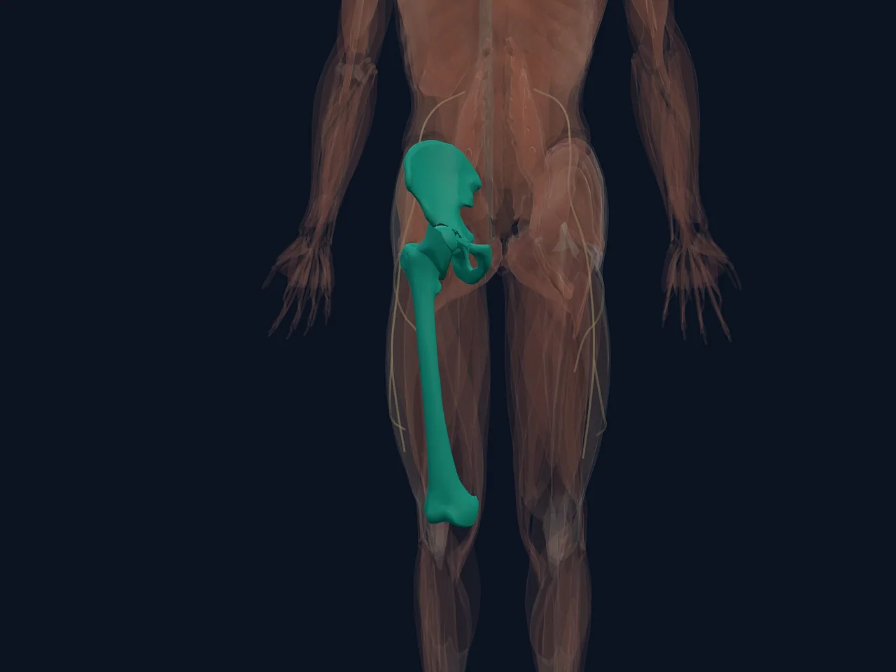
The hip joint is one of the body's largest joints and one of its most stable. It is a ball-and-socket joint: the round head of the thigh bone (femoral head) sits inside a cup-shaped socket (acetabulum) formed by the pelvis.
That deep socket gives the hip a wide range of motion while still carrying body weight during walking, running, lifting and climbing stairs. Understanding how the joint is built helps explain why hip problems often show up as groin pain, outer-hip pain or stiffness.
Location and Connections
- The socket is part of the pelvis, on each side of the lower torso
- The ball is the upper end of the femur (thigh bone)
- A ring of tough tissue called the labrum lines the rim of the socket
- Strong ligaments wrap around the joint capsule and limit extreme motion
- Large muscles surround the joint: the gluteals behind, the hip flexors in front, and the adductors along the inner thigh
What It Does
The hip transmits weight from the upper body to the legs, allows the leg to move forward, backward, sideways and in rotation, and absorbs force during impact. Its design favours stability: the socket is deep, the capsule is strong, and the surrounding muscles add dynamic support.
The labrum works like a gasket, deepening the socket, sealing in joint fluid and helping distribute load. The smooth articular cartilage covering the ball and socket lets the bones glide with very little friction.
Common Problems
- Hip osteoarthritis — gradual wearing of joint cartilage, more common with age
- Hip labral tears — injury to the rim of the socket, often with twisting or deep hip positions
- Femoroacetabular impingement (FAI) — bony shape changes that cause pinching during movement
- Hip flexor strain or irritation — front-of-hip or groin pain, common in runners and dancers
- Gluteal tendinopathy and trochanteric pain — outer-hip pain from the tendons on the side of the hip
- Referred pain from the lower back — hip-area pain that actually starts in the spine
Pain location gives clues but does not identify the exact source by itself. Groin pain often points toward the hip joint itself, while outer-hip pain more often involves the tendons beside the joint.
Clinical Relevance
Assessment matters when hip pain follows significant trauma, comes with an inability to bear weight, or includes deformity, fever, or sudden severe pain. Sudden inability to move the hip normally, or pain with numbness or weakness in the leg, also needs prompt evaluation.
Related Anatomy
- Gluteal muscles — the large muscles behind and beside the hip
- Lumbar spine — a common source of referred hip-area pain
References
- StatPearls. "Anatomy, Bony Pelvis and Lower Limb, Hip Joint." https://www.ncbi.nlm.nih.gov/sites/books/NBK526019/
- StatPearls. "Anatomy, Bony Pelvis and Lower Limb, Hip Joint." https://pubmed.ncbi.nlm.nih.gov/29262200/