Overview
Femoroacetabular impingement syndrome (FAI syndrome) — often called hip impingement — is a pattern of hip or groin pain in which symptoms, clinical signs and a particular hip shape fit together. It is not diagnosed from an X-ray or MRI alone. Many people have cam or pincer shape features and never develop symptoms.
The hip is a ball-and-socket joint. In some hips, the ball is less round at the head-neck junction (cam morphology) or the socket provides more coverage at the rim (pincer morphology). During flexion and rotation — such as a deep squat, a low seat or a cut and turn — these shapes can contribute to a painful pinch and may load the labrum or cartilage. Hip strength, movement options, training load, cartilage health and the person’s goals also influence symptoms.
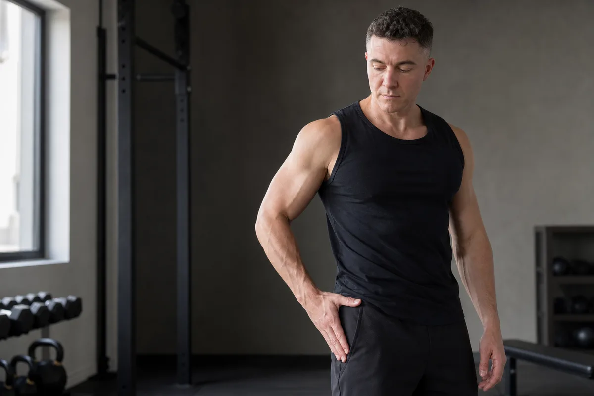
Anatomy: Where Does Hip Impingement Happen?
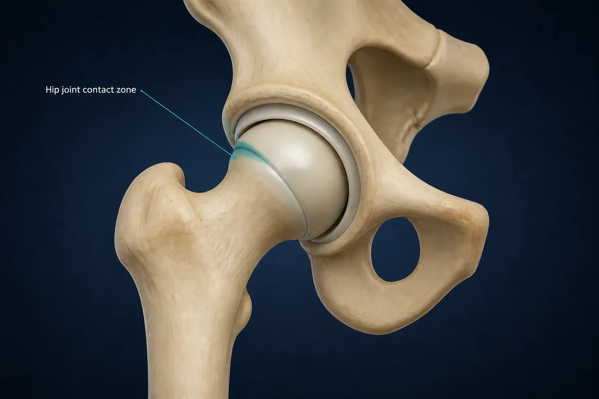
The rounded femoral head sits inside the acetabulum (hip socket). The labrum is a fibrocartilage rim around the socket; articular cartilage covers the joint surfaces.
- Cam morphology: extra bone or reduced roundness around the femoral head-neck junction. It can contact the front of the socket during flexion and rotation.
- Pincer morphology: greater socket coverage in a particular area. It may compress the labrum at the rim in certain positions.
- Labrum and cartilage: may be irritated or damaged when joint mechanics and repeated loading do not match the hip’s current capacity.
Words such as “bump,” “lesion” or “deformity” can sound alarming. A shape feature is common and does not by itself mean that a hip is damaged or needs treatment.
Typical Pattern
- Deep pain at the front of the hip or in the groin; outer-hip or buttock pain can occur but is less specific
- A sharp pinch with a deep squat, low chair, stair climb, turn, pivot or twist
- A dull ache after sitting, driving or working with the hip bent for a long time
- Stiffness, reduced comfortable flexion or a sense that the hip does not move smoothly
- Clicking or catching can be present, but painless clicking alone does not diagnose FAI syndrome
- Symptoms in activities that require repeated deep range, rotation, kicking, skating or cutting
FAI syndrome is more commonly discussed in active younger and middle-aged adults, but age or sport alone does not establish the diagnosis.
Why It Develops
Cam morphology commonly develops during skeletal growth and is seen more often in some athletic populations. Pincer morphology describes socket coverage and may be focal or global. Neither finding guarantees pain or arthritis.
Symptoms can become noticeable when a hip is repeatedly asked to tolerate more deep range, rotation, force or volume than it currently can. This may follow a training change, a sport season, a return after time off or no clear single event. Exercise does not “cause” the bony shape, but demanding activity may expose a mismatch between capacity and load.
What Else Can Feel Similar?
Groin and front-of-hip pain have many possible causes. A thorough assessment considers:
- Hip labral tear: can coexist with FAI syndrome and create painful catching or locking
- Hip flexor or adductor strain: often more local and provoked by resisted muscle testing or a rapid stretch
- Hip osteoarthritis: more common with joint-space change, persistent stiffness and progressive activity limits
- Gluteal tendinopathy: usually causes more lateral hip pain and may hurt when lying on that side
- Femoral-neck stress injury: deep weight-bearing pain that worsens with walking, running or hopping; needs prompt assessment
- Lumbar spine, nerve or abdominal-wall causes: may include back pain, tingling, pain down the leg or symptoms with coughing and trunk effort
Do not self-diagnose a deep hip pinch from a single test or scan report.
How It Is Diagnosed
A clinician assesses the onset, precise pain location, training demands, effect of sitting and sport, hip and spine movement, strength and symptom-provoking positions. A flexion-adduction-internal-rotation position is commonly used, but no single test confirms that FAI syndrome is the pain source.
- X-rays: generally assess bone shape, socket coverage and signs of arthritis first
- MRI without contrast or MR arthrogram: may assess labrum, cartilage, bone and other soft tissue when X-rays are negative or nondiagnostic and the clinical question remains
- Diagnostic injection: can help show whether pain is coming from inside the hip joint; temporary relief supports an intra-articular source but does not identify one structure with certainty
Current imaging guidance considers both MRI without contrast and MR arthrography appropriate next studies when impingement or dysplasia is suspected after nondiagnostic radiographs.
Treatment
Treatment normally starts with a non-surgical plan that fits the person’s symptoms and goals:
- Activity adjustment: modify painful deep flexion, pivoting and prolonged sitting without abandoning all movement
- Progressive strength: build gluteal, adductor, hip-rotator and trunk strength in ranges the hip tolerates
- Movement retraining: adjust squat depth, stance, lifting strategy, sitting setup or sport technique if a movement clearly reproduces symptoms
- Conditioning: retain tolerable cardiovascular work and general strength to avoid unnecessary deconditioning
- Pain relief: short-term medication or an injection may be considered with a qualified clinician when appropriate; relief should support rehabilitation rather than replace it
There is no single “best” physiotherapy recipe or calendar. A staged progression, with the response later that day and the next morning guiding advancement, is usually more useful than repeatedly pushing through a sharp pinch.
Rehabilitation: A Staged Approach
-
Settle the pinch
- Modify sharp-pinch positions
- Break up long periods of hip-flexed sitting
Progress when: Daily movement is settling without a sharp flare
-
Restore control
- Strengthen glutes, adductors and hip rotators
- Build trunk control in comfortable ranges
Progress when: There is no sharp pain or next-day escalation
-
Build capacity
- Progress squat, hinge and single-leg loading
- Add rotation and relevant range gradually
Progress when: Strength matches the demands of daily life or training
-
Return to demand
- Run → accelerate → decelerate → change direction
- Restore deep range, kicking or pivoting progressively
Progress when: There is no later or next-morning flare
Stage 1: Settle the Irritable Pattern
- Reduce the depth, load, speed or frequency of positions that cause a sharp pinch
- Break up long sitting and find a hip angle that is more comfortable
- Keep walking and general activity within a tolerable range
- Avoid repeatedly “testing” the painful end-range position
Stage 2: Rebuild Control and Baseline Strength
- Train gluteals, adductors, hip rotators and trunk control in comfortable ranges
- Use bridges, hinges, step patterns and isometrics as tolerated
- Restore movement options gradually without forcing a painful end point
- Keep fitness through activities that do not meaningfully flare symptoms
Stage 3: Build Strength for Life, Lifting or Sport
- Progress squat, split-squat, hinge and single-leg loading in relevant ranges
- Reintroduce rotation and deeper range only as symptoms and control permit
- Build capacity for the person’s work, running, skating, kicking or change-of-direction demands
- Change one major variable at a time: range, load, speed or volume
Stage 4: Return to Demanding Activity
Progress from steady conditioning to acceleration, deceleration, planned cuts, pivots, kicking or deep-range lifting. For lifters, depth and load can be restored gradually; for field and court athletes, high-speed change of direction should be prepared before full competition.
When Surgery Is Considered
Hip arthroscopy may be discussed when a well-matched rehabilitation trial has not restored acceptable function and the history, examination and imaging support a treatable intra-articular problem. Surgery may reshape symptomatic cam or pincer morphology and repair or address labral or cartilage damage when appropriate.
It is a shared decision rather than an automatic next step from an X-ray. Cartilage health, arthritis, dysplasia or instability, the person’s goals and the match between symptoms and imaging all influence the potential benefit. Arthroscopy does not guarantee a perfect hip or eliminate every source of groin pain.
Recovery Expectations
Nonsurgical Care
Some people notice better sitting tolerance and daily function within weeks; restoring sport-specific strength and confidence often takes longer. Progress depends on irritability, training demands, movement goals and whether a labral or cartilage problem also contributes. Reassess the diagnosis and plan when a sensible, progressive programme is not moving things forward.
After Arthroscopy
Recovery depends on the procedures performed. Weight-bearing, range restrictions, brace use and return-to-running timelines vary, especially if labral repair, cartilage work or bone reshaping was performed. Follow the surgeon’s protocol instead of a generic online calendar. A return to unrestricted sport is generally measured in months rather than weeks.
Return-to-Sport Checklist
Before unrestricted training or competition, aim for:
- Daily activity and prolonged sitting without meaningful symptom escalation
- Hip motion adequate for the activity without a sharp pinch or apprehension
- Hip and trunk strength appropriate for the task and close to the other side when side-to-side comparison is relevant
- Controlled squat, hinge, landing and single-leg patterns
- Gradual tolerance of running, acceleration, deceleration and planned change of direction where needed
- Sport-specific kicking, pivoting, skating or deep range at increasing intensity
- No significant flare later that day or the following morning
When to Get Assessed
- Inability to bear weight or a major limp after trauma
- A hip that is locked and cannot move through its usual range
- Severe or rapidly worsening pain, especially at rest or at night
- Fever, chills, redness, swelling or feeling systemically unwell
- Deep groin pain that worsens with walking or hopping, which can indicate a bone stress injury
- Numbness, progressive weakness, bowel or bladder changes, or pain travelling down the leg
- Persistent symptoms that do not respond to sensible load adjustment and rehabilitation
Common Questions
Does hip impingement always need surgery?
No. A diagnosis of FAI syndrome does not automatically mean surgery. Many people first use education, load adjustment and progressive rehabilitation. Surgery is considered when symptoms remain limiting and the complete clinical picture supports a treatable joint problem.
Can I keep squatting or playing sport?
Often, yes—with temporary adjustments. A narrower depth, altered stance, lighter load, different exercise variation or reduced frequency may help you stay active while rebuilding capacity. The long-term goal is usually to restore the movement needed for your life or sport rather than to avoid it forever.
Does an X-ray bump mean my hip is damaged?
No. Cam and pincer shape features are common in people without symptoms. FAI syndrome requires symptoms, clinical signs and imaging findings that fit together.
Can stretching fix a hip pinch?
Forcing a hard, painful stretch into a pinching position is rarely useful. Some people benefit from comfortable mobility work, but strength, load management and movement options generally matter more than chasing maximum range.
Related Reading
References
- StatPearls. "Femoroacetabular Impingement." https://www.ncbi.nlm.nih.gov/books/NBK547699/
- AAOS OrthoInfo. "Femoroacetabular Impingement." https://www.orthoinfo.org/diseases--conditions/femoroacetabular-impingement/
- Griffin DR, et al. "The Warwick Agreement on femoroacetabular impingement syndrome (FAI syndrome): an international consensus statement." British Journal of Sports Medicine. 2016. https://doi.org/10.1136/bjsports-2016-096743
- American College of Radiology. "ACR Appropriateness Criteria: Chronic Hip Pain." Revised 2022. https://acsearch.acr.org/docs/69425/Narrative/
- Kemp JL, et al. "The 2019 International Society of Hip Preservation physiotherapy agreement on assessment and treatment of femoroacetabular impingement syndrome." Journal of Hip Preservation Surgery. 2021. https://pmc.ncbi.nlm.nih.gov/articles/PMC8349584/
This information is for education only and is not a medical diagnosis. InjuryAtlas does not replace an assessment by a qualified health professional. If you have severe pain, visible deformity, numbness, loss of function, or symptoms that are getting worse, seek medical care promptly.