Overview
A hip labral tear affects the acetabular labrum — the ring of fibrocartilage around the rim of the hip socket. The labrum helps deepen the socket, maintain a fluid seal, distribute load and support joint stability. When it is irritated or torn, some people develop deep groin pain, painful clicking or difficulty with pivoting and deep hip positions.
The scan finding and the pain experience are not the same thing. Labral changes can appear on imaging in people without symptoms, while hip impingement, dysplasia, cartilage damage, tendons, the groin and the lower back can create a similar pain pattern. A useful diagnosis connects the history, examination and imaging instead of treating the MRI report alone.
Many symptomatic labral tears begin with nonsurgical care. Surgery may be considered when a well-matched rehabilitation trial has not restored acceptable function and the labrum or underlying hip shape remains a plausible source of symptoms.
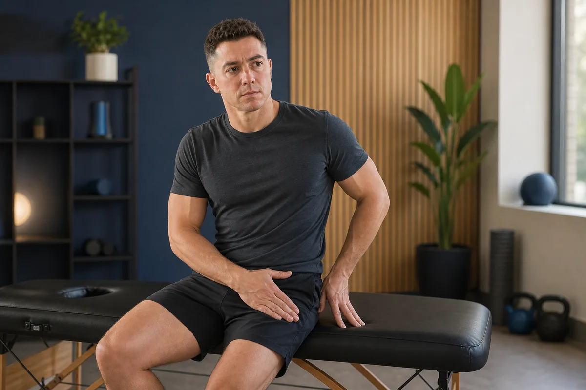
Anatomy: What Does the Labrum Do?
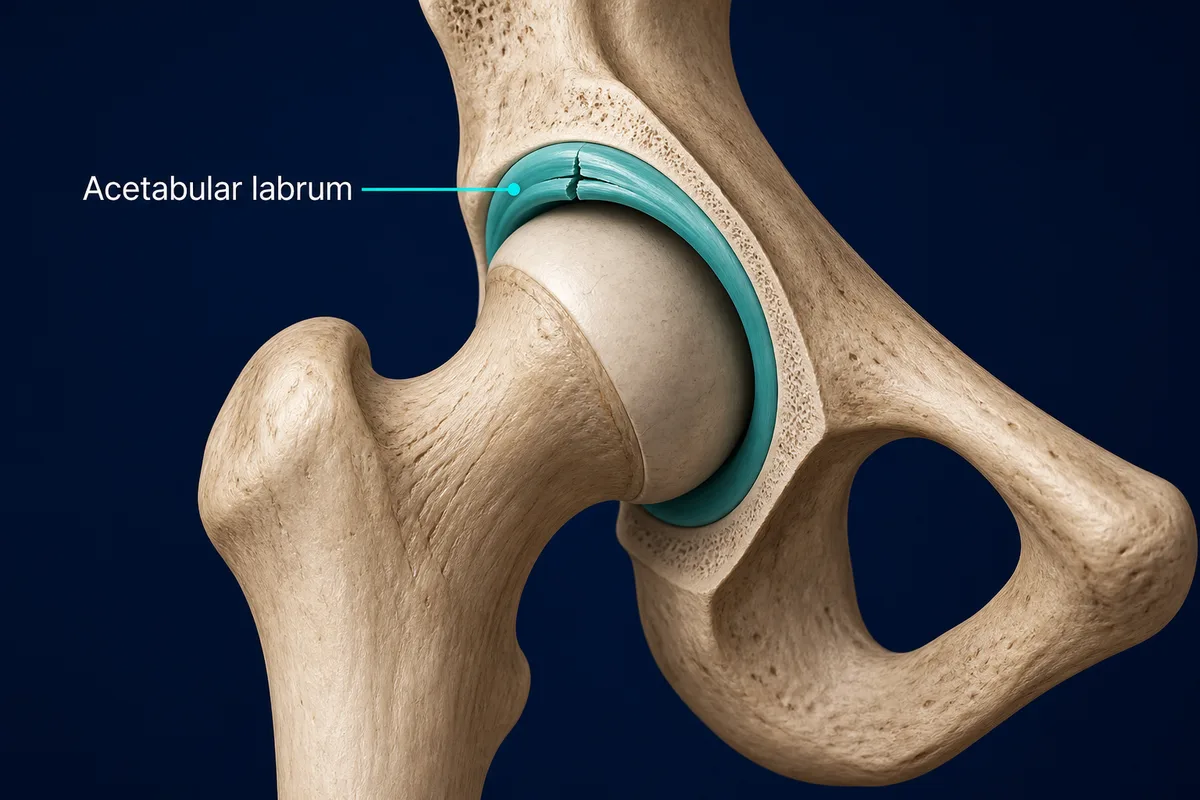
The hip is a ball-and-socket joint. The rounded femoral head sits inside the acetabulum, while the labrum attaches around most of the socket rim.
- Joint seal: helps retain fluid between the joint surfaces and contributes to smooth load transfer
- Socket depth and stability: increases the effective depth of the acetabulum and supports control of the femoral head
- Load distribution: helps spread pressure around the rim of the socket
- Sensation: contains nerve fibres, so injury or compression can contribute to pain
The labrum has a limited blood supply, especially along parts of its inner edge. Tear location, tissue quality, cartilage health and the shape of the hip all influence whether a tear is repairable and whether it is likely to be clinically important.
Tear Patterns and Associated Hip Shape
A labral tear may be traumatic, repetitive or degenerative. Common descriptions include:
- Anterior or anterosuperior tear: the most commonly discussed location, often stressed during hip flexion and rotation
- Detachment: the labrum pulls away from the socket rim
- Intrasubstance or degenerative tear: tissue frays or splits within the labrum
- Complex tear: more than one tear pattern is present
The tear often coexists with another structural feature:
- Cam morphology: extra bone at the femoral head-neck junction can contact the socket rim during flexion and rotation
- Pincer morphology: greater socket coverage can compress the labrum
- Acetabular dysplasia: a shallower socket may place greater demand on the labrum for stability
- Cartilage damage or osteoarthritis: changes the prognosis and may reduce the benefit expected from arthroscopy
These shapes are not automatically diseases. They matter when they fit the symptoms and examination.
Typical Pattern
- Deep pain at the front of the hip or in the groin; buttock or outer-hip pain is less specific
- Pain with pivoting, cutting, twisting, deep squatting or rising from a low seat
- Discomfort after prolonged sitting or driving with the hip bent
- Painful clicking, catching or locking during movement
- Reduced hip motion or a pinching sensation near the end of flexion
- Symptoms during sport that involves repeated rotation, kicking or deep hip range
Clicking alone does not diagnose a tear. Many hips click without pain, and snapping tendons outside the joint can create a similar sensation.
Why It Develops
The labrum can become symptomatic through:
- Repeated compression or shear: deep flexion, rotation and high training volume may repeatedly load the socket rim
- Femoroacetabular impingement syndrome: symptoms, examination findings and cam or pincer morphology occur together
- Trauma: a fall, tackle, collision, forceful twist or hip dislocation
- Dysplasia or instability: a shallow socket or excessive motion can increase labral demand
- Degeneration: tissue quality and cartilage health can change with age and joint disease
- Rapid workload change: a sudden increase in cutting, kicking, skating, dance or deep-range lifting can exceed current capacity
What Else Can Feel Similar?
Deep hip or groin pain has several possible sources:
- Femoroacetabular impingement syndrome: often overlaps with labral injury and may be the main mechanical driver
- Hip osteoarthritis: stiffness, reduced motion and load-related pain, particularly when joint-space loss is present
- Hip flexor or adductor injury: pain is often more reproducible with resisted muscle testing or local palpation
- Gluteal tendinopathy: pain is usually more lateral and may be aggravated by lying on the affected side
- Femoral-neck stress injury: deep weight-bearing pain, sometimes progressing to rest or night pain; prompt assessment is important
- Inguinal or abdominal-wall injury: symptoms may increase with coughing, sneezing, sit-ups or forceful trunk effort
- Lumbar spine or nerve-related pain: back pain, numbness, tingling or symptoms travelling down the leg
True locking, inability to bear weight, worsening night pain or systemic symptoms should not be managed as a routine labral tear without assessment.
How It Is Diagnosed
A clinician reviews the onset, exact pain location, training demands, clicking or locking and the effect of sitting, walking and rotation. Examination commonly includes gait, hip and spine motion, strength, palpation and symptom-provoking positions. No single physical test can confirm that a labral tear is the pain source.
Imaging is interpreted in stages:
- Radiographs: usually assess joint space, arthritis, dysplasia and cam or pincer morphology first
- MRI without contrast: can show the labrum, cartilage, bone and surrounding soft tissues
- MR arthrogram: places contrast inside the joint and can better define some labral tears, although modern high-quality MRI may already be informative
- Image-guided anaesthetic injection: may help determine how much pain is coming from inside the joint; temporary relief supports an intra-articular source but does not identify the labrum with certainty
When radiographs are negative or nondiagnostic and a labral tear is suspected, both MRI without intravenous contrast and MR arthrography are considered appropriate next studies in current imaging guidance.
Nonsurgical Treatment
Initial care aims to reduce irritation while rebuilding capacity around the hip:
- Load adjustment: temporarily reduce painful deep flexion, forceful pivoting and repeated end-range positions without stopping all activity
- Education: identify which positions are provocative and vary sitting, training depth and weekly workload
- Progressive strength: train gluteals, adductors, hip rotators and trunk control, then reintroduce demanding ranges
- Movement retraining: adjust squat depth, cutting mechanics or sport technique when these clearly reproduce symptoms
- Conditioning: maintain tolerable cardiovascular work and general strength so the entire programme does not become deconditioning
- Medication or injection: may be considered with a qualified clinician when appropriate; symptom relief should support rehabilitation rather than replace it
A systematic review of non-arthritic hip-related pain found that just over half of participants reported a satisfactory response to nonsurgical care, but the studies included several related diagnoses and did not establish one superior physiotherapy recipe. The practical lesson is to use an individualized, progressive programme and reassess the diagnosis when progress stalls.
Rehabilitation: A Staged Approach
-
Reduce irritability
- Keep comfortable daily movement
- Modify painful deep flexion and pivoting
Progress when: Sitting and walking are settling
-
Restore control
- Strengthen glutes, adductors and hip rotators
- Build trunk control in comfortable ranges
Progress when: There is no sharp pain or next-day flare
-
Build capacity
- Progress squats, hinges and single-leg loading
- Add controlled rotation and conditioning
Progress when: Strength and range approach the other side
-
Return to sport
- Run → accelerate → decelerate → change direction
- Add kicking, pivoting and deep range gradually
Progress when: There is no later or next-morning escalation
Stage 1: Reduce Irritability Without Complete Rest
- Shorten or modify deep squats, low sitting and twisting that cause a sharp pinch
- Keep walking and daily activity within a tolerable range
- Avoid repeatedly testing the most painful position
- Maintain comfortable upper-body and opposite-limb training
Stage 2: Restore Control and Baseline Strength
- Train trunk and pelvic control
- Strengthen gluteals, adductors and hip rotators in comfortable ranges
- Use controlled bridge, hinge, step and isometric patterns as tolerated
- Restore hip range gradually without forcing a hard, painful end point
Stage 3: Build Strength Through Relevant Range
- Progress squats, split squats, hinges and single-leg loading
- Increase hip strength in the ranges required for work or sport
- Add controlled rotation and change-of-direction preparation
- Monitor the response later that day and the following morning
Stage 4: Return to Running, Cutting or Deep-Range Sport
Progress from straight-line conditioning to acceleration, deceleration, direction change and sport-specific drills. Lifters and dancers can gradually restore depth and rotation. Increase one major variable at a time—range, load, speed or volume.
When Surgery Is Considered
Hip arthroscopy may be discussed when:
- Symptoms continue to limit valued activity after a structured nonsurgical programme
- The history, examination, imaging and sometimes injection all support an intra-articular pain source
- A repairable labral lesion or associated impingement can be addressed
- Joint cartilage and overall hip shape make a meaningful benefit reasonably likely
The operation may repair the labrum, reconstruct deficient tissue or selectively trim unstable tissue. Surgeons may also reshape symptomatic cam or pincer morphology. The plan must account for dysplasia and instability because removing bone or labral tissue without addressing the overall mechanics can be unhelpful.
Arthroscopy is not a guaranteed cure for every MRI tear. More advanced osteoarthritis, substantial cartilage loss and poorly matched symptoms can reduce the likelihood of a strong result.
Recovery Expectations
Nonsurgical Care
A focused rehabilitation trial commonly runs for at least several weeks, with progress judged by sitting tolerance, daily function, strength and the ability to reintroduce sport-specific load. Some people improve quickly; longstanding symptoms, dysplasia, significant impingement or cartilage damage may require a longer plan or specialist review.
After Arthroscopy
Recovery depends on whether the labrum was repaired, reconstructed or debrided and whether bone or cartilage procedures were added. Early weight-bearing, range restrictions and brace use vary substantially between surgical protocols. Follow the surgeon's procedure-specific plan rather than a generic online calendar.
Return to unrestricted sport is usually measured in months rather than weeks. Published protocols vary, and readiness should be based on healing restrictions, strength, range, impact tolerance, cutting or rotation capacity and sport-specific testing.
Return-to-Sport Checklist
Before unrestricted training or competition, aim for:
- Daily activity and prolonged sitting without meaningful symptom escalation
- Hip range adequate for the sport without a sharp pinch or apprehension
- Hip and trunk strength close to the other side and appropriate for the activity
- Controlled squat, hinge, landing and single-leg patterns
- Running, acceleration, deceleration and planned cutting completed progressively
- Sport-specific rotation, kicking or deep range tolerated at increasing intensity
- No significant flare later that day or the following morning
- Confidence in the positions and movements the sport requires
Seek Urgent or Prompt Assessment
- Inability to bear weight or a major limp after trauma
- A hip that is locked and cannot move through its usual range
- Severe or rapidly worsening pain, especially at rest or at night
- Fever, chills, redness, swelling or feeling systemically unwell
- Deep groin pain that worsens with walking or hopping, raising concern about bone stress injury
- Numbness, progressive weakness, bowel or bladder changes, or pain travelling down the leg
- A new groin bulge with severe pain, nausea or vomiting
- Persistent symptoms that are not responding to sensible load adjustment and rehabilitation
Common Questions
Does a hip labral tear always need surgery?
No. Many people begin with education, load adjustment and progressive rehabilitation. Surgery is a shared decision for symptoms that remain limiting when the clinical picture and imaging support a treatable intra-articular problem.
Can a labral tear heal on its own?
The labrum has limited blood supply, so the tissue may not return to a completely normal appearance. Symptoms and function can still improve substantially as irritation settles and strength, movement options and load tolerance improve.
Is clicking proof of a labral tear?
No. Painless clicking is common, and tendons outside the joint can snap. Painful catching or locking deserves assessment, but the diagnosis requires the full clinical picture.
Should I avoid squats forever?
Usually not. Early modifications to depth, stance, load or frequency may help. Rehabilitation can then restore the ranges required for daily life, lifting or sport when symptoms permit.
Which scan is best?
Radiographs usually assess bone shape and arthritis first. MRI without contrast and MR arthrography are both appropriate options when a labral tear remains suspected after nondiagnostic radiographs. The choice depends on local imaging quality, the clinical question and specialist preference.
Related Reading
- Hip joint anatomy
- Femoroacetabular impingement
- Hip osteoarthritis
- Groin pain
- General hip loading principles
References
- American College of Radiology. "ACR Appropriateness Criteria: Chronic Hip Pain." Revised 2022. https://acsearch.acr.org/docs/69425/Narrative/
- Probst DT, et al. "What is the rate of response to non-operative treatment for hip-related pain? A systematic review with meta-analysis." Journal of Orthopaedic & Sports Physical Therapy. 2023. https://pmc.ncbi.nlm.nih.gov/articles/PMC10176100/
- Arakgi ME, et al. "Management of Labral Tears in the Hip: A Consensus Statement." 2025. https://pmc.ncbi.nlm.nih.gov/articles/PMC11758551/
- Petchprapa CN, et al. "Imaging of the acetabular labrum." Seminars in Musculoskeletal Radiology. 2013. https://pubmed.ncbi.nlm.nih.gov/23787979/
- Grzybowski JS, et al. "Rehabilitation Following Hip Arthroscopy — A Systematic Review." 2015. https://pmc.ncbi.nlm.nih.gov/articles/PMC4443726/
This information is for education only and is not a medical diagnosis. InjuryAtlas does not replace an assessment by a qualified health professional. If you have severe pain, visible deformity, numbness, loss of function, or symptoms that are getting worse, seek medical care promptly.