Overview
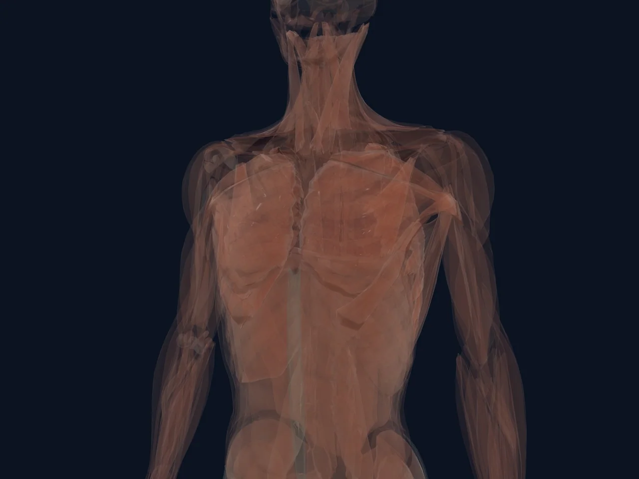
The intercostal muscles are the thin muscles that fill the spaces between the ribs. They sit in three layers and work with the diaphragm to expand and recoil the ribcage with every breath.
They are also a common site of "pulled" chest muscles — sharp pain between the ribs that is worse with breathing, coughing, twisting or reaching.
Location and Connections
- Three layers — external, internal and innermost intercostals — between each pair of ribs
- The external intercostals lie most superficially and mainly help lift the ribcage during inhalation
- The internal and innermost intercostals lie deeper and mainly assist forced exhalation
- Nerves and blood vessels run in grooves along the underside of each rib, protected by the rib bone above
What It Does
The intercostals move the ribs during breathing, help stabilize the chest during forceful movements, and assist with twisting and bending of the torso. During heavy breathing, coughing or sneezing, they work much harder — which is why strains hurt most in those moments.
Common Problems
- Intercostal strain — a pulled or overstretched intercostal muscle, often after coughing, twisting, lifting or a direct blow
- Pain between the ribs with deep breaths, coughing or reaching
- Muscle spasm and tenderness over the affected space
- Intercostal irritation that can feel similar to rib fracture or costochondritis
Intercostal pain is common and usually settles with load adjustment, but chest pain can also be cardiac or pulmonary — heavy, pressing pain with shortness of breath or pain spreading to the arm or jaw needs urgent assessment.
Clinical Relevance
Because the intercostals work with every breath, they cannot be completely rested. Recovery involves managing pain, keeping breathing normal, and gradually returning to twisting, lifting and reaching. Persistent pain, difficulty breathing or pain after trauma deserves assessment.
Related Anatomy
References
- StatPearls. "Anatomy, Thorax, Muscles." https://www.ncbi.nlm.nih.gov/sites/books/NBK538321/