Overview
The femoral nerve is the largest branch of the lumbar plexus. It is formed mainly from the L2-L4 nerve roots, travels through the pelvis beside the psoas and iliacus, then passes beneath the inguinal ligament into the front of the thigh.
Anatomy at a Glance
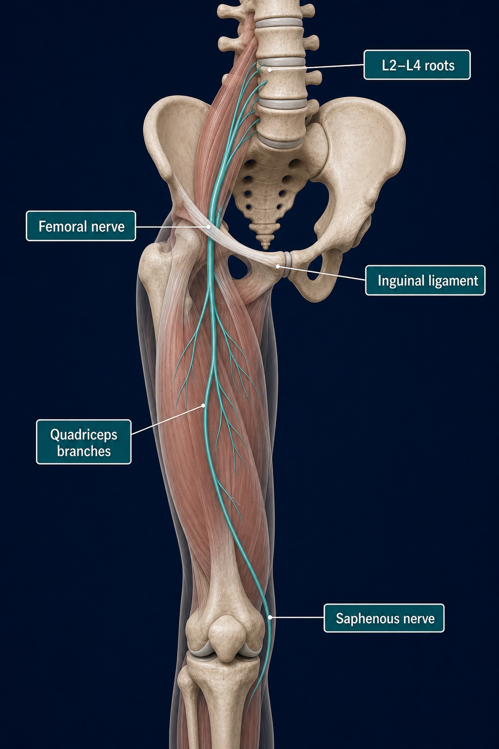
The main nerve passes beneath the inguinal ligament before dividing into branches for the quadriceps and sensation. Its saphenous branch continues toward the inner knee and lower leg.
Key Role
- Powers the quadriceps for straightening the knee
- Supplies parts of the iliacus, sartorius and pectineus
- Carries sensation from the front of the thigh
- Continues through the saphenous nerve to supply the inner lower leg and foot
- Sends sensory branches to the hip and knee joints
Route and Connections
The nerve forms within the psoas region, emerges between psoas major and iliacus, and enters the thigh just outside the femoral artery. It then divides into several motor and sensory branches rather than staying as one large cord.
How It Is Usually Loaded
Normal movement does not “stretch” the femoral nerve in isolation. The nerve and its surrounding tissues must slide as the hip and knee move. Prolonged hip extension, swelling, trauma or pressure around the pelvis and groin can change that movement or irritate the nerve.
Watch Out For
Femoral nerve problems are uncommon compared with muscle or joint pain. New quadriceps weakness, repeated knee buckling, reduced knee reflexes or spreading numbness across the front thigh and inner leg need assessment. Symptoms after pelvic surgery, a major fall or bleeding risk deserve prompt attention.
Connected Guides
References
- StatPearls. "Anatomy, Bony Pelvis and Lower Limb: Thigh Femoral Nerve." https://www.ncbi.nlm.nih.gov/books/NBK556065/
This information is for education only and is not a medical diagnosis.