Overview
The femur is the long bone of the thigh. Its rounded head fits into the hip socket, its shaft carries load through the thigh, and its lower end helps form the knee.
It is built for high forces, but different parts fail in different ways. A fall can fracture the upper femur, while repeated impact can create a bone stress injury at the femoral neck or shaft.
Anatomy at a Glance
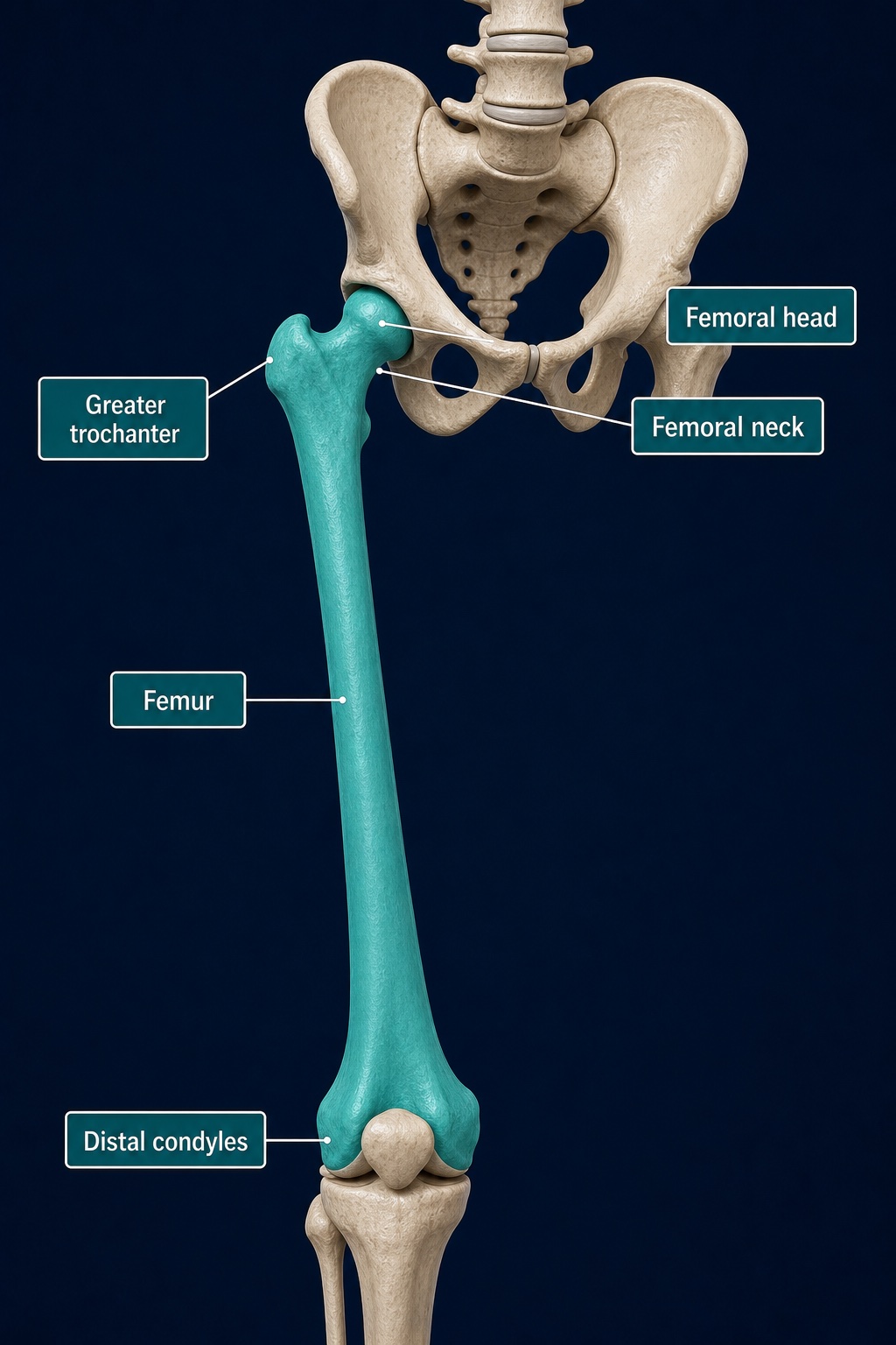
The femoral head and neck form the upper end of the bone, while the distal condyles help form the knee. The greater trochanter is the prominent outer-hip attachment point.
Key Landmarks
- Femoral head: the rounded surface inside the hip socket
- Femoral neck: the narrow bridge between the head and shaft
- Greater trochanter: the large outer-hip attachment point for gluteal muscles and tendons
- Lesser trochanter: the smaller inner landmark where the iliopsoas attaches
- Shaft: the long central section surrounded by the thigh muscles
- Condyles: the rounded lower surfaces that meet the tibia at the knee
Key Role
The femur transfers force between the pelvis and lower leg. Its shape gives the hip a large range of motion while providing broad attachment sites for the glutes, adductors, quadriceps, hamstrings and deep hip muscles.
How It Is Usually Loaded
Walking and stairs repeatedly load the femur through body weight and muscle pull. Running, jumping and heavy lower-body lifts increase those forces. Bone normally adapts to gradual loading, but sudden training increases, inadequate recovery or reduced bone health can outpace that adaptation.
Watch Out For
Deep groin or thigh pain that becomes more focal with impact should not automatically be treated as a muscle strain. Inability to stand after a fall, a shortened or rotated leg, or severe groin pain requires urgent assessment. Runners with progressively worsening weight-bearing pain also need prompt evaluation for bone stress injury.
Connected Guides
References
- StatPearls. "Anatomy, Bony Pelvis and Lower Limb: Femur." https://www.ncbi.nlm.nih.gov/books/NBK532982/
This information is for education only and is not a medical diagnosis.