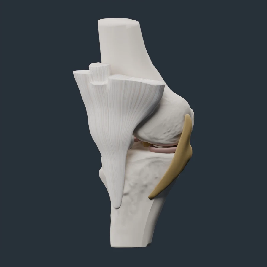
Anatomy at a Glance
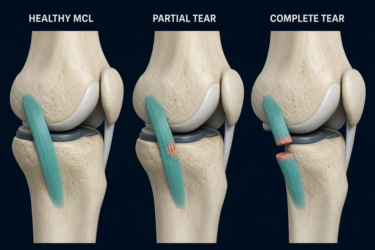
The MCL is the broad ligament along the inner knee. Sprains range from partial fibre disruption to a complete loss of continuity.
Overview
An MCL sprain damages the broad ligament complex along the inner knee. It commonly happens when a force to the outside of the knee drives the knee inward, although a twist or awkward ski fall can also injure it.
What It Usually Feels Like
- Pain and tenderness along the inner knee
- Swelling that may be more localized than with an ACL tear
- Pain when the knee is stressed sideways
- Stiffness or difficulty walking comfortably
- Instability in a higher-grade injury
Inner joint-line pain can also come from the medial meniscus or joint surface. Pain location alone is not a diagnosis.
What the Grades Mean
- Grade 1: fibres are stretched or mildly damaged, without meaningful laxity
- Grade 2: partial tear with increased looseness but a remaining endpoint
- Grade 3: complete tear with substantial laxity
Pain intensity does not reliably tell you the grade. A clinician assesses tenderness and stability at different knee angles and checks for associated ACL or meniscus injury.
Treatment
Most isolated MCL injuries begin without surgery. Early care may include temporary protection, a hinged brace when indicated, restoring motion and progressive strengthening. Complete rest for weeks is rarely the whole plan; the knee usually needs a graded return to movement and load.
Surgery may be considered for selected complete tears, certain avulsions, persistent instability or combined ligament injuries. The exact tear location and associated damage matter.
When to Get Assessed
Arrange an assessment if the knee feels unstable, swelling is substantial, weight bearing is difficult, motion is limited or the injury involved a forceful collision. A deformity, numbness, colour change or locked knee needs urgent care.
Related Reading
References
- AAOS OrthoInfo. “Collateral Ligament Injuries.” https://orthoinfo.aaos.org/en/diseases--conditions/collateral-ligament-injuries/
- Encinas-Ullán CA, Rodríguez-Merchán EC. “Isolated medial collateral ligament tears: an update on management.” https://pmc.ncbi.nlm.nih.gov/articles/PMC6129956/
This information is for education only and is not a medical diagnosis. InjuryAtlas does not replace an assessment by a qualified health professional. If you have severe pain, visible deformity, numbness, loss of function, or symptoms that are getting worse, seek medical care promptly.