Anatomy at a Glance
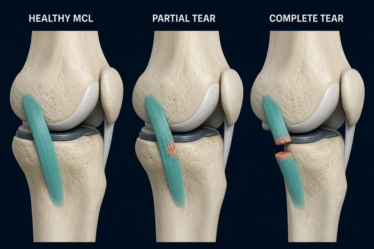
The MCL is the broad ligament along the inner knee. Sprains range from partial fibre disruption to a complete loss of continuity.
Start With the Injury You Actually Have
An MCL injury does not automatically mean surgery or months without using the leg. The medial collateral ligament runs along the inside of the knee, and many isolated sprains heal with protection followed by progressive movement, strength and sport-specific loading.
The details still matter. A mild sprain with no looseness is not managed exactly like a complete tear, an avulsion, a combined ACL/MCL injury or a ligament that has been surgically repaired. Before copying an exercise plan, find out:
- whether the injury is isolated or other structures are involved;
- whether the knee is stable during ordinary walking;
- whether a hinged brace or crutches were prescribed;
- whether knee motion or weight bearing is restricted; and
- what activities you need to return to.
This guide focuses mainly on non-operative rehabilitation for an isolated MCL sprain. If you had surgery or injured the ACL, meniscus or another ligament, follow the restrictions from your surgical and rehabilitation team.
Before You Begin
Pain on the inside of the knee does not confirm an MCL tear. The medial meniscus, joint surface, pes anserine tendons and other tissues can hurt in a similar area. The MCL injury guide explains the usual injury pattern and assessment.
Arrange a prompt assessment when:
- the knee feels markedly loose or repeatedly gives way;
- swelling is large or developed rapidly after the injury;
- you cannot bear weight or walk several steps;
- the knee is locked and cannot fully straighten;
- there was a high-force collision, dislocation or obvious deformity;
- the foot becomes cold, pale, weak or numb; or
- calf swelling, chest pain or shortness of breath develops.
Why MCL Rehabilitation Is Different From ACL Rehabilitation
The MCL has a comparatively good blood supply and many isolated injuries can heal without reconstruction. Early rehabilitation therefore balances two goals: protect the healing ligament from excessive inward stress while preventing unnecessary stiffness, weakness and loss of confidence.
That does not mean testing the ligament every day. Sideways stress, cutting and awkward twisting usually return later than comfortable straight-line walking and controlled strength work.
A hinged knee brace is sometimes used for moderate or high-grade injuries. It can limit unwanted sideways movement while allowing bending and straightening. Bracing is not needed for every sprain, and the setting and duration should come from a clinician who has assessed the knee.
Phase 1: Protect the Knee and Settle the Irritation
Main goals: protect the healing ligament, reduce pain and swelling, regain comfortable straightening, begin bending and activate the thigh muscles.
Early management may include:
- a hinged brace or crutches when prescribed;
- walking only as far as you can without a marked limp;
- comfortable knee-straightening practice;
- heel slides for controlled bending;
- quadriceps tightening and straight-leg raises when tolerated;
- ankle pumps and calf raises; and
- gentle hip strengthening that does not pull the knee inward.
Keep the knee aligned over the foot during walking and exercise. Avoid deliberately pushing the knee inward to “stretch” the MCL. The ligament does not need repeated valgus stress to prove that it is healing.
Cold packs, compression and elevation may help short-term comfort or swelling. They are optional symptom tools, not the main treatment.
Signs you may be ready to progress
- Pain and swelling are trending down.
- The knee can straighten fully or is steadily approaching the other side.
- Bending is improving without a large flare afterward.
- You can tighten the quadriceps and raise the straight leg without it sagging.
- Walking is becoming more even within your brace and weight-bearing instructions.
Phase 2: Restore Walking and Basic Strength
Main goals: walk normally, recover knee motion, rebuild thigh and hip strength and improve balance.
Common building blocks include:
- sit-to-stand;
- shallow supported squats;
- low step-ups;
- bridges and hamstring curls;
- calf raises;
- stationary cycling when bending is comfortable; and
- supported balance that progresses toward single-leg standing.
Start in a range where the knee stays controlled. The goal is not to keep every exercise easy; it is to add resistance without reproducing instability or repeatedly increasing medial-knee pain and swelling.
As walking normalizes, gradually increase distance before adding hills, speed or unpredictable ground. If a brace was prescribed, do not stop using it simply because pain has eased. Pain often improves before the ligament is ready for contact or cutting.
Signs you may be ready to progress
- Full or nearly full knee motion.
- Normal walking without a limp.
- Little or no reactive swelling after exercise.
- Controlled sit-to-stand, step-up and shallow single-leg tasks.
- No episodes of giving way.
- Medial tenderness and pain are steadily improving.
Phase 3: Rebuild Single-Leg Capacity
Main goals: develop stronger single-leg control, tolerate heavier loading and prepare for impact.
This phase may include:
- progressively loaded squats and leg press;
- split squats and reverse lunges;
- higher step-ups and controlled step-downs;
- hamstring, quadriceps and calf strengthening;
- single-leg balance with reaching or light external challenge; and
- lateral hip strength to help control the thigh and pelvis.
Do not judge readiness from one exercise. The leg needs to tolerate repeated work without the knee drifting inward, pain building through the session or swelling appearing later.
For everyday activity, this phase may be enough to resume longer walks, stairs and a gradual return to physical work. Running, jumping and cutting require another layer of preparation.
Phase 4: Return to Running and Low-Level Impact
Main goals: tolerate repeated impact, restore running capacity and introduce controlled jumping and landing.
Useful checkpoints before running include:
- full knee motion;
- no more than minimal swelling;
- comfortable brisk walking;
- no instability;
- good control during repeated single-leg squats or step-downs; and
- sufficient strength on objective testing for the planned activity.
Begin with short walk-jog intervals on a predictable surface. Increase total running time before adding speed, hills or sharp direction changes.
Jumping can start with two-leg take-offs and landings, then progress toward single-leg and multi-direction work. The knee should remain aligned and confident rather than collapsing inward or avoiding load on the recovering side.
Phase 5: Reintroduce Cutting, Contact and Sport
Main goals: restore acceleration, braking, direction change, reactive movement and confidence under realistic demands.
Later rehabilitation may include:
- faster running and sprint exposure;
- planned direction changes before reactive cutting;
- lateral shuffles and crossover steps;
- repeated jumping, landing and hopping;
- sport- or job-specific drills under fatigue; and
- gradual contact exposure when relevant.
Straight-line running does not prove that the MCL is ready for an opponent, an unexpected collision or a hard plant-and-cut. Reintroduce predictable drills first, then speed, reaction and contact as separate progressions.
A brace may be recommended for part of the return to sport in selected higher-grade injuries. This is an individual decision, not a requirement for every athlete.
How Long Does MCL Rehabilitation Take?
MCL recovery varies with injury grade, tear location, stability, associated injuries and activity demands. A mild isolated sprain may return to activity within a few weeks, while a higher-grade injury commonly needs a longer period of protection and progressive rehabilitation. Surgery or a combined ligament injury follows a substantially different timeline.
Published rehabilitation programs vary considerably, and research does not support one precise schedule for every isolated MCL injury. Use time as context—not as automatic clearance.
More useful progression markers include:
- full motion;
- no meaningful swelling;
- no tenderness that is worsening;
- a stable clinical examination;
- strength and hop performance appropriate to the task;
- confident running, cutting and landing mechanics; and
- completion of practice or work progressions without a reaction.
What Should Exercise Feel Like?
Muscle effort and mild temporary discomfort can be acceptable. A session is more likely to be appropriate when:
- pain remains mild rather than climbing with every repetition;
- the knee does not buckle or feel loose;
- movement stays controlled;
- swelling does not meaningfully increase; and
- walking and knee motion are not worse the next day.
Sharp medial pain, a sense of separation or giving way, or a repeated next-day flare means the current range, load, speed or volume needs to be reconsidered.
Common MCL Rehabilitation Mistakes
Assuming every inside-knee injury is an MCL sprain
The medial meniscus and other structures can produce similar pain. A forceful injury or persistent symptoms deserve assessment.
Stopping all movement for too long
Protection can be necessary, but prolonged unnecessary immobilization may contribute to stiffness and weakness. Follow the brace and movement plan given for the injury grade.
Testing sideways stress too early
Repeatedly pushing the knee inward does not speed healing. Build controlled strength first and reintroduce lateral movement progressively.
Returning when pain is gone but control is not
Low pain during daily life does not prove readiness for cutting, skiing, contact or uneven ground.
Treating a combined injury like an isolated MCL sprain
ACL, meniscus, posteromedial-corner and multiligament injuries can change protection, surgical decisions and rehabilitation timing.
When Surgery Changes the Plan
Most isolated MCL injuries begin with non-operative care, including many complete tears. Surgery may be considered for selected avulsions, entrapment, persistent instability, chronic laxity or combined ligament injuries.
After repair or reconstruction, the surgeon may restrict weight bearing, brace settings, knee bending or hamstring loading. A generic non-operative program should not override those instructions.
Questions to Ask Your Clinician
- Is this an isolated MCL injury?
- What grade and location is the tear?
- Do I need a hinged brace, and when can it be unlocked or removed?
- Are there weight-bearing or range-of-motion restrictions?
- What findings will determine when I can run?
- How will strength, stability and cutting ability be tested?
- Do I need a brace when I return to sport or work?
Related InjuryAtlas Resources
- MCL sprain and tear
- MCL anatomy
- ACL vs MCL tear
- ACL rehabilitation guide
- General knee loading principles
- Medial meniscus
- Explore the MCL in 3D
References
- Svantesson E, et al. “Shedding light on the non-operative treatment of the forgotten side of the knee: rehabilitation of medial collateral ligament injuries—a systematic review.” BMJ Open Sport & Exercise Medicine. 2024. https://pmc.ncbi.nlm.nih.gov/articles/PMC11202733/
- Sanford Health. “Rehabilitation of Isolated MCL Knee Sprains: Rehabilitation Guideline.” https://www.sanfordhealth.org/-/media/org/files/medical-professionals/resources-and-education/isolated-mcl-sprain.pdf
- Massachusetts General Brigham Sports Medicine. “Rehabilitation after Injury to the Medial Collateral Ligament of the Knee.” https://www.massgeneral.org/assets/mgh/pdf/orthopaedics/sports-medicine/physical-therapy/rehabilitation-protocol-for-medial-collateral-ligament-injury-mcl.pdf
- Chahla J, et al. “The posteromedial corner of the knee: an international expert consensus statement on diagnosis, classification, treatment, and rehabilitation.” Knee Surgery, Sports Traumatology, Arthroscopy. 2021. https://pmc.ncbi.nlm.nih.gov/articles/PMC7586411/
This information is for education only and is not a medical diagnosis or personalised rehabilitation program. Follow the restrictions from your clinician after a high-grade injury or operation. Seek medical care promptly for a locked knee, major trauma, repeated giving way, inability to bear weight, deformity, numbness or colour change, worsening calf swelling, chest pain or shortness of breath.