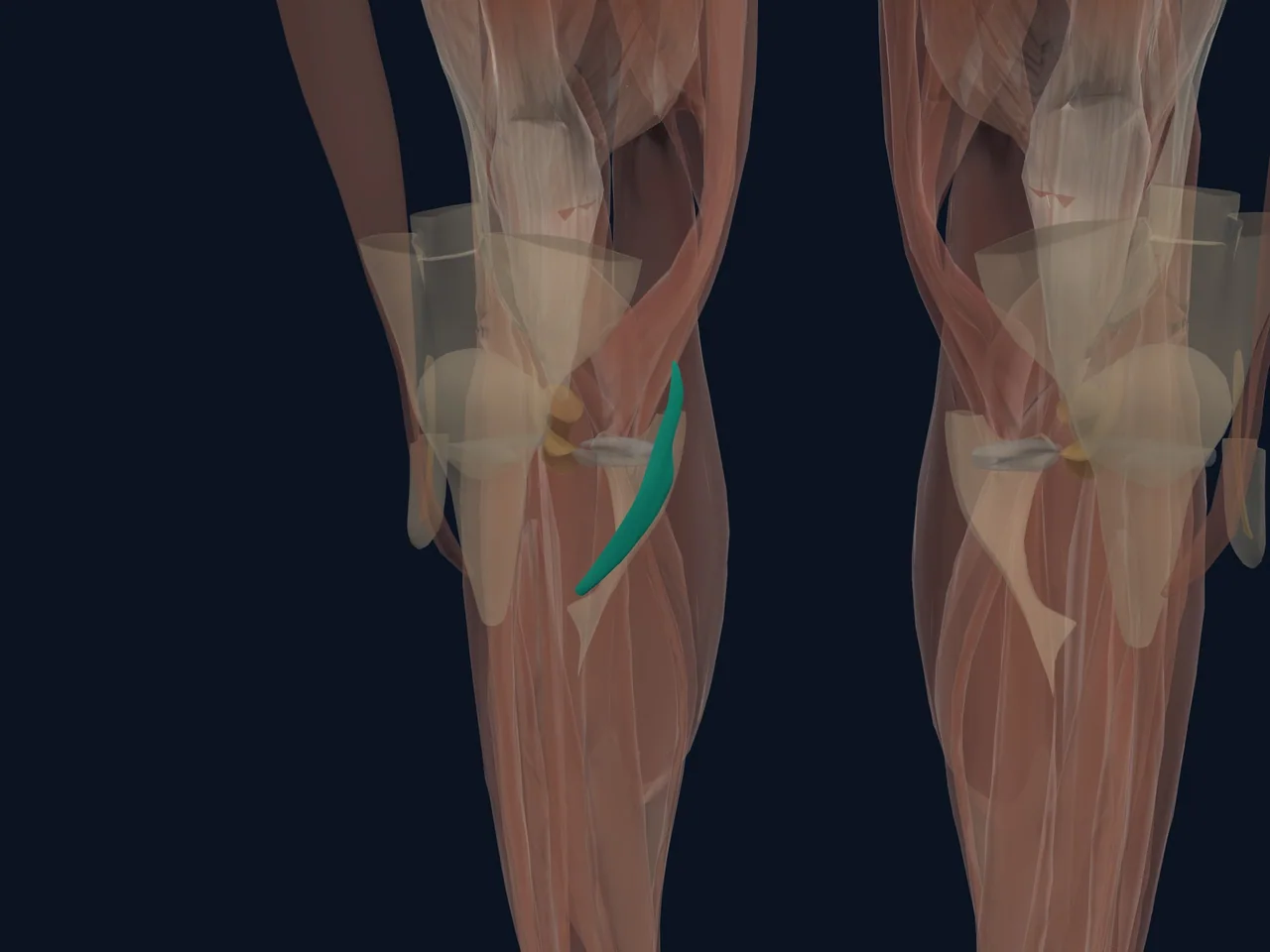
Overview
The medial collateral ligament (MCL) is the broad ligament complex along the inside of the knee. It is a primary restraint against valgus force—the knee moving inward relative to the thigh and lower leg—and also contributes to rotational control.
Location and Attachments
- Runs from the inner femur to the inner tibia
- Includes superficial and deep components
- Deep fibres connect with the joint capsule and medial meniscus
- Works with the cruciate ligaments and surrounding muscles
What It Does
The MCL stabilizes the medial knee during walking, cutting and contact. Its broad shape and layered attachments let it resist different forces across the knee's range of motion.
Common Injury Pattern
An MCL sprain often follows a force to the outside of the knee that drives it inward. Non-contact twisting can also injure it. Pain and tenderness commonly occur along the inner knee, but medial meniscus and joint injuries can feel similar.
Clinical Relevance
MCL sprains are graded by tissue damage and laxity, but a grade cannot be determined from pain intensity alone. Assessment is especially important after substantial trauma, marked instability, rapid swelling or inability to bear weight because more than one structure may be involved.
Related Anatomy
References
- StatPearls. "Anatomy, Bony Pelvis and Lower Limb, Knee Medial Collateral Ligament." https://www.ncbi.nlm.nih.gov/books/NBK507780/
- AAOS OrthoInfo. "Collateral Ligament Injuries." https://orthoinfo.aaos.org/en/diseases--conditions/collateral-ligament-injuries/