Overview
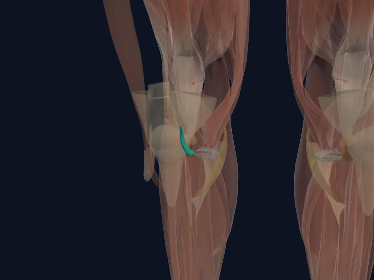
The anterior cruciate ligament (ACL) is one of two crossing ligaments deep inside the knee. It joins the femur to the tibia and is a major restraint against the shinbone sliding too far forward. It also contributes to rotational control during cutting, pivoting and landing.
Location and Attachments
- Runs through the centre of the knee joint
- Attaches on the lateral femoral condyle and the front portion of the tibial plateau
- Crosses in front of the PCL
- Works with the menisci, collateral ligaments, muscles and joint capsule
What It Does
The ACL resists excessive forward translation of the tibia relative to the femur and helps control rotation. Its bundles change tension as the knee bends, which lets the ligament contribute across a range of positions rather than acting like a simple fixed rope.
Common Injury Pattern
ACL injury often occurs during a rapid change of direction, awkward landing or sudden deceleration. Contact injuries also occur. A pop, fast swelling or a feeling that the knee gave way may raise concern, but none of those signs proves a tear by itself.
ACL injuries can occur with damage to the medial meniscus, lateral meniscus, cartilage or other ligaments.
Clinical Relevance
Diagnosis combines the mechanism, examination and—when appropriate—imaging. Treatment decisions depend on instability, associated damage, sport or work demands, age and personal goals. Not every ACL injury follows the same path.
Related Anatomy
- Posterior cruciate ligament
- Medial collateral ligament
- Medial meniscus
- Lateral meniscus
- ACL rehabilitation guide
References
- StatPearls. "Anatomy, Bony Pelvis and Lower Limb, Knee Anterior Cruciate Ligament." https://www.ncbi.nlm.nih.gov/books/NBK559233/
- AAOS. "Clinical Practice Guideline: Management of Anterior Cruciate Ligament Injuries." https://www.aaos.org/quality/quality-programs/lower-extremity-programs/anterior-cruciate-ligament-injuries/