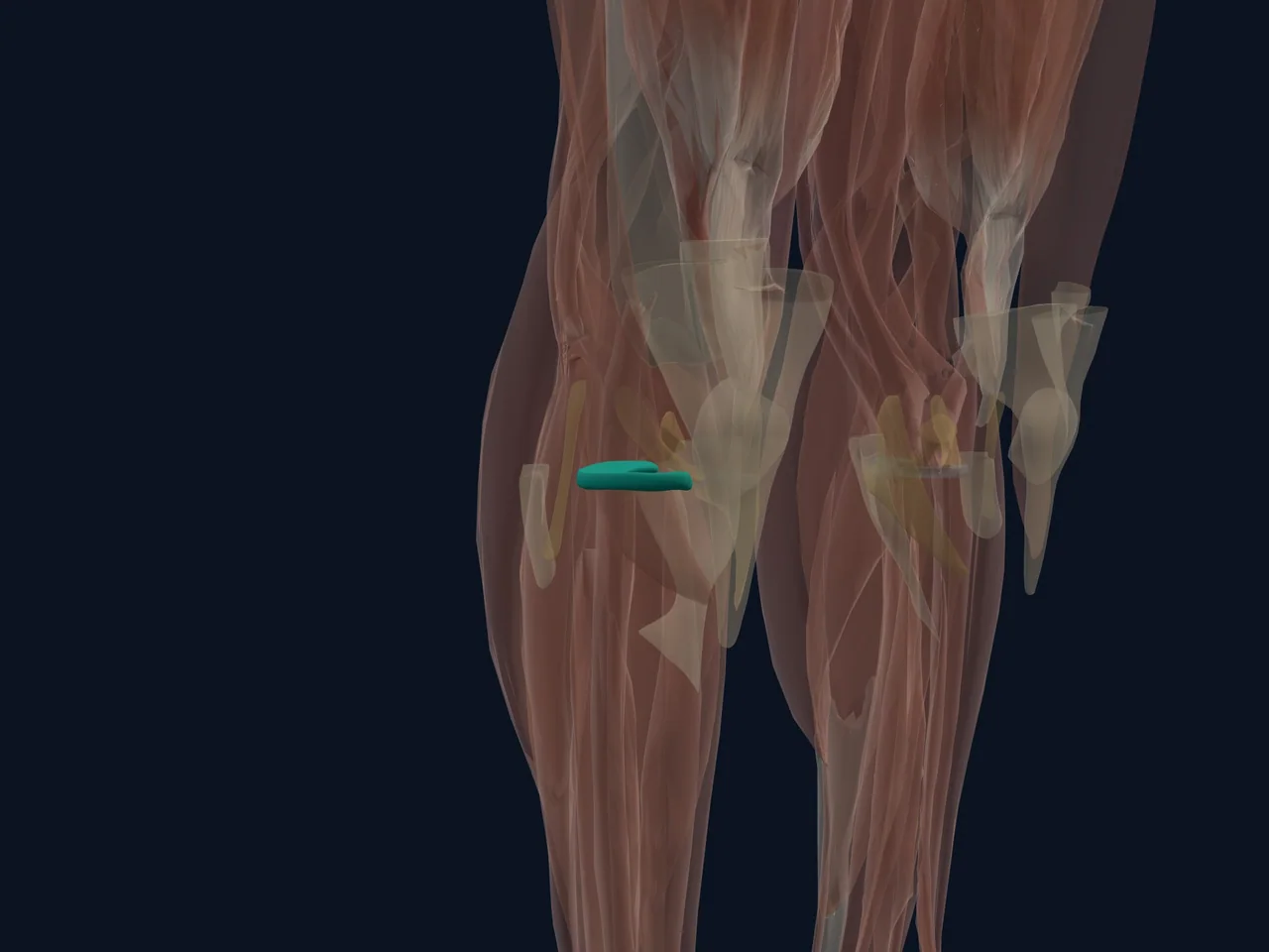
Overview
The lateral meniscus is the rounded, crescent-like pad of fibrocartilage between the femur and tibia on the outside of the knee. It distributes load, absorbs shock, improves joint fit and helps the knee move smoothly.
Location and Attachments
- Sits on the lateral tibial plateau
- Anchors to the tibia through anterior and posterior horns
- Has a gap near the popliteus tendon that allows more movement
- Does not attach directly to the LCL
- Is generally more mobile than the medial meniscus
What It Does
The lateral meniscus carries a substantial part of the load through the outer knee. Its wedge shape deepens the relatively flat tibial surface and improves joint congruence. It also supports lubrication, proprioception and secondary stability.
Blood Supply and Healing
Only the peripheral portion has a meaningful direct blood supply; the inner portion is relatively avascular. Tear location affects healing potential, but it is not the only factor. Tear shape, stability, tissue quality, symptoms and activity goals all influence management.
Common Problems
- Traumatic tear during loaded rotation
- Tear associated with ACL injury
- Degenerative tear
- Meniscal cyst related to a horizontal tear
Outer joint-line pain can also come from the LCL, iliotibial band, cartilage or proximal tibiofibular joint.
Clinical Relevance
A scan should be interpreted alongside symptoms and examination because meniscal changes can exist without being the main pain source. True locking after injury, rapid swelling or inability to bear weight deserves assessment.
Related Anatomy
- Meniscus rehabilitation guide
- Medial meniscus
- Lateral collateral ligament
- Anterior cruciate ligament
References
- StatPearls. "Anatomy, Bony Pelvis and Lower Limb, Knee Lateral Meniscus." https://www.ncbi.nlm.nih.gov/books/NBK519494/
- StatPearls. "Knee Meniscal Tears." https://www.ncbi.nlm.nih.gov/books/NBK431067/