Anatomy at a Glance
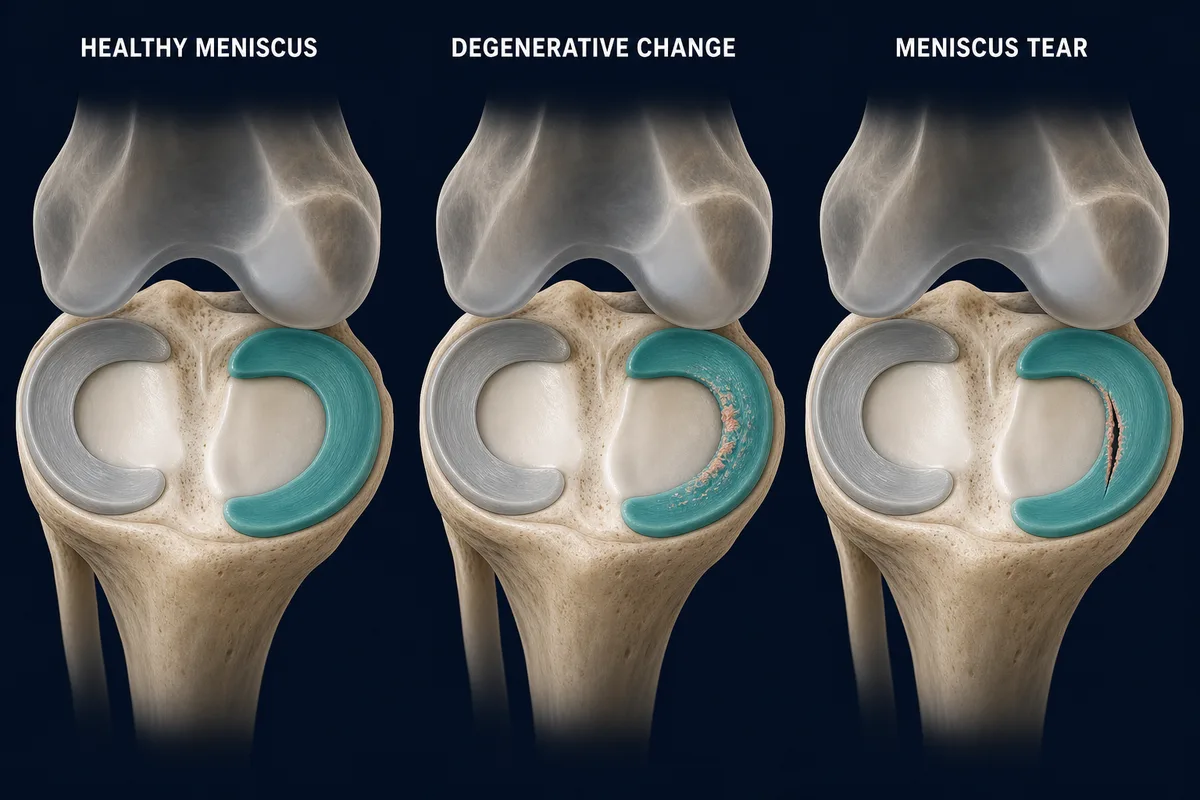
Meniscal changes range from gradual fraying to a defined tear. The appearance of a tear does not by itself determine whether it is painful or needs surgery.
First, Find Out Which Meniscus Pathway You Are On
The word meniscus tear does not describe one recovery plan. A small, non-displaced tear managed without surgery may be loaded according to symptoms and function. A partial meniscectomy usually permits a comparatively quick return to motion and weight bearing. A repaired meniscus needs time for the tissue to heal, and a root, radial or complex repair may require substantially more protection.
Before following a program, clarify:
- whether the tear is traumatic, degenerative, displaced or associated with arthritis;
- whether the medial or lateral meniscus is involved;
- the tear pattern and location;
- whether other structures, such as the ACL or cartilage, were injured;
- whether surgery removed damaged tissue or repaired it; and
- whether your surgeon restricted weight bearing, knee bending, squatting or twisting.
This guide explains the shared rehabilitation principles and the important differences between pathways. It cannot replace the restrictions given after an operation.
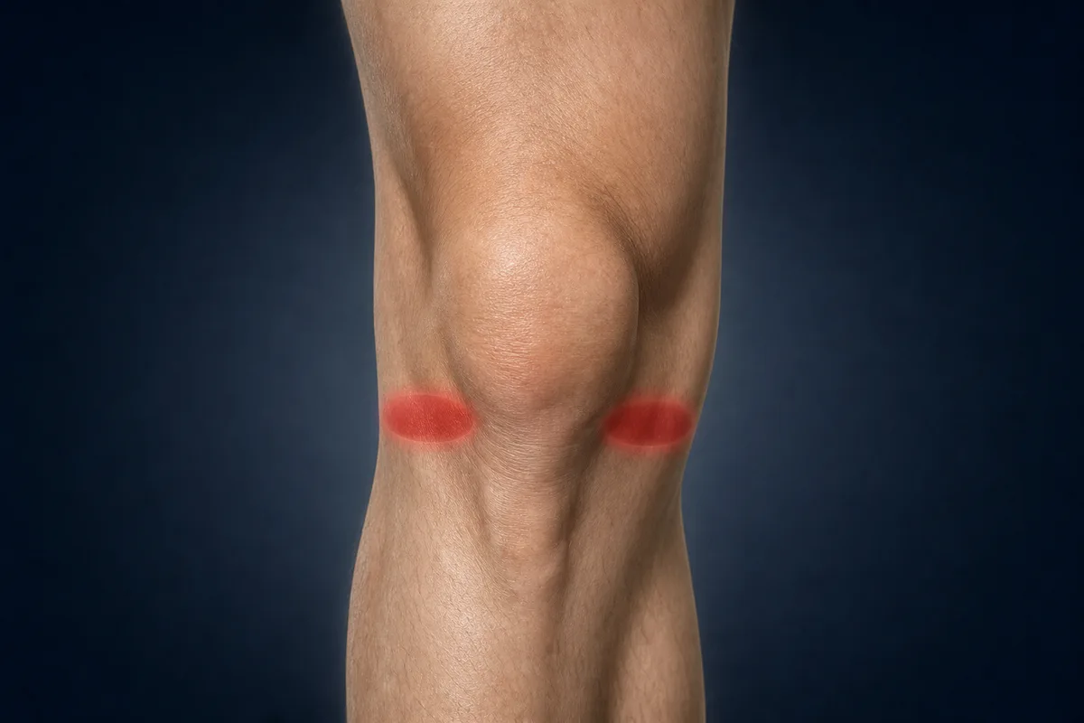
Meniscus-related pain is often felt along the inner or outer joint line, but location alone cannot confirm a tear.
Before You Begin
Joint-line pain, clicking or pain with squatting does not confirm a meniscus tear. Meniscal findings are also common on MRI in people without symptoms. The scan, injury history, examination and behaviour of the knee need to make sense together.
Arrange a prompt assessment when:
- the knee is genuinely locked and cannot fully straighten;
- a traumatic twist caused marked swelling or inability to bear weight;
- the knee repeatedly gives way;
- pain or swelling is steadily worsening rather than settling;
- the knee or leg looks deformed;
- the foot becomes cold, pale or numb; or
- after surgery, the wound becomes increasingly red, hot or draining, or you develop fever, calf swelling, chest pain or shortness of breath.
True locking is different from a knee that is painful or stiff. A displaced tear can physically block movement and deserves timely assessment.
Four Common Rehabilitation Pathways
1. Non-operative rehabilitation
Many non-displaced traumatic tears and many degenerative meniscus symptoms can begin with rehabilitation. The goal is not to make an MRI finding disappear. It is to restore motion, strength and tolerance for the activities that matter to you.
Research does not support automatically sending every meniscus tear to surgery. Symptoms that persist despite a well-performed rehabilitation trial, repeated mechanical locking and certain repairable or displaced tears may change the discussion.
2. Partial meniscectomy
During partial meniscectomy, unstable tissue is trimmed rather than repaired. Because there is no repaired tear to protect, weight bearing and motion commonly progress as tolerated. Rehabilitation is generally criterion-based and often advances faster than after repair.
The knee can still become swollen and the quadriceps can still weaken. “Nothing was stitched” does not mean strength and impact capacity return immediately.
3. Meniscus repair
A repair preserves meniscal tissue but needs biological healing. Progression should account for the tear pattern, repair stability and surgeon's instructions. Some repairs permit early weight bearing and motion; others restrict them.
Rehabilitation after repair uses both time and milestones. Feeling good early does not mean the repair is ready for deep loaded bending, pivoting or running.
4. Root, radial or complex repair
Root and complete radial tears disrupt the meniscus's ability to transmit load through circumferential fibres. Their rehabilitation is often more conservative than a stable vertical repair, with specific restrictions on weight bearing, knee flexion and loaded squatting.
Do not copy a standard meniscectomy or uncomplicated repair protocol after one of these procedures.
Phase 1: Settle the Knee and Restore Basic Control
Main goals: protect the injured or repaired tissue, reduce swelling, regain comfortable extension, begin bending within restrictions and activate the quadriceps.
Early work may include:
- crutches or a brace when prescribed;
- short, even walking within weight-bearing instructions;
- comfortable knee-straightening practice;
- heel slides within the permitted range;
- quadriceps tightening;
- straight-leg raises when the knee does not sag;
- ankle pumps and calf work; and
- gentle hip strengthening.
After repair, do not force bending because the knee feels stiff. Some surgeons limit flexion during the early healing period, particularly after root, radial or complex repairs.
Cold packs, compression and elevation can help short-term comfort and swelling. They are optional symptom tools, not substitutes for a progressive rehabilitation plan.
Signs you may be ready to progress
- Swelling is small and trending down.
- The knee reaches full extension or is steadily approaching it.
- Bending is improving within your restrictions.
- You can perform a straight-leg raise without lag.
- Walking is becoming more even within the prescribed weight-bearing level.
- Pain and joint-line irritation are not escalating after each session.
Phase 2: Recover Walking, Motion and Everyday Strength
Main goals: normalize walking, restore motion, rebuild quadriceps and hip strength and regain control during everyday tasks.
Common exercises include:
- sit-to-stand;
- shallow supported squats when permitted;
- low step-ups;
- bridges and hamstring work;
- calf raises;
- stationary cycling once adequate bending is available; and
- balance work that progresses toward one leg.
Start with a range the knee tolerates. Deeper flexion increases compression on the back of the meniscus, and loaded deep squatting may be restricted after repair. Depth is a progression—not a test you need to pass immediately.
After partial meniscectomy or during non-operative care, progression can usually follow symptoms and function more closely. After repair, the calendar and surgical restrictions still matter even when exercises feel easy.
Signs you may be ready to progress
- Full or nearly full motion for your stage.
- Normal walking without a marked limp.
- Little or no reactive swelling after exercise.
- Controlled sit-to-stand, step-up and shallow single-leg tasks.
- Improving strength without repeated joint-line flares.
- No locking or giving-way episodes.
Phase 3: Rebuild Single-Leg Strength and Deeper Range
Main goals: restore lower-limb strength, tolerate more demanding knee angles and prepare for impact or heavier work.
This phase may include:
- progressively loaded squats and leg press;
- split squats and reverse lunges;
- higher step-ups and controlled step-downs;
- knee-extension and hamstring strengthening;
- hip and calf strengthening; and
- single-leg balance with reaching or external challenge.
Increase depth gradually. A knee that tolerates a shallow squat may react when depth, load and volume all rise together. Change one variable at a time so you can understand the response.
After repair, confirm when loaded flexion beyond 90 degrees, deep squatting and kneeling are allowed. The answer depends on the tear and operation, not simply how many weeks have passed.
Phase 4: Return to Running and Impact
Main goals: tolerate repeated impact, restore running capacity and introduce jumping and landing.
Useful checkpoints before running include:
- full active knee motion;
- no more than minimal swelling;
- comfortable brisk walking;
- good control during repeated single-leg squats or step-downs;
- strength appropriate to the planned activity; and
- clearance of any repair-specific time restriction.
Begin with short walk-jog intervals on a predictable surface. Increase total running time before speed, hills or direction changes.
Jumping typically progresses from two-leg take-offs and landings toward single-leg and multi-direction tasks. Pain or swelling that accumulates later that day or the next morning is a reason to adjust the dose.
Phase 5: Return to Pivoting, Sport and Demanding Work
Main goals: restore speed, braking, rotation, reaction and confidence under realistic demands.
Later rehabilitation may include:
- faster running and sprint exposure;
- planned direction changes before reactive cutting;
- lateral movement and crossover steps;
- repeated jumping, landing and hopping;
- gradual exposure to kneeling or deep work positions; and
- sport- or job-specific tasks under fatigue.
Straight-line jogging does not prove readiness for a planted twist, an opponent, uneven ground or repeated deep squatting. Build predictable tasks first, then speed, reaction and fatigue.
Return-to-sport decisions should combine symptoms, motion, swelling, strength, movement control, activity-specific testing and confidence. One good training session is not the same as being prepared for full competition.
How Long Does Meniscus Rehabilitation Take?
The pathway matters more than the label alone.
- Non-operative care: progress is based mainly on symptom response and functional milestones. A meaningful trial commonly lasts several months.
- Partial meniscectomy: return to activity is often measured in weeks, but swelling, strength and sport demands determine the actual pace.
- Meniscus repair: vertical repairs generally require at least several months of rehabilitation. Return to pivoting sport is commonly later.
- Root, radial, horizontal or complex repair: rehabilitation may extend six to nine months or longer depending on healing and activity demands.
These ranges are context, not promises. The 2024 international consensus recommends criterion-based progression after meniscectomy and combined time- and criterion-based progression after repair.
What Should Exercise Feel Like?
Muscle effort and mild temporary discomfort can be acceptable. A session is more likely to be appropriate when:
- pain stays mild rather than rising with each repetition;
- movement remains controlled;
- the knee does not catch, lock or buckle;
- swelling does not meaningfully increase; and
- walking and motion are not worse the following day.
Joint-line pain that becomes sharper as depth or twisting increases is useful feedback. Reduce range, load, speed or volume instead of repeatedly forcing through it.
Common Meniscus Rehabilitation Mistakes
Treating every tear the same
A degenerative MRI finding, a stable vertical tear, a displaced bucket-handle tear and a root repair have different implications.
Assuming surgery automatically means faster recovery
Partial meniscectomy often progresses quickly; repair deliberately progresses more slowly to preserve tissue.
Testing deep squats and twisting too early
These movements can be reintroduced, but repeatedly testing the most provocative position is not a rehabilitation strategy.
Resting until all pain disappears
Avoiding every activity can leave the leg weaker and less prepared. Most pathways need progressive loading within appropriate restrictions.
Ignoring swelling
The knee may feel acceptable during exercise and swell later. Track the response that evening and the next morning.
Following a generic protocol after repair
The operative report and surgeon's restrictions matter. Root and radial repairs may need different protection from simpler repairs.
When to Return to Your Clinician
Arrange reassessment when:
- the knee becomes locked or repeatedly catches with loss of motion;
- swelling keeps returning despite sensible load adjustments;
- extension is not improving;
- the knee repeatedly gives way;
- symptoms are steadily worsening;
- progress has stalled despite a consistent rehabilitation trial; or
- postoperative pain, swelling or wound symptoms are outside the expectations given by your surgical team.
Questions to Ask After Meniscus Surgery
- Was the meniscus repaired or partially removed?
- Which meniscus and which tear pattern were involved?
- Was it a root, radial, horizontal, complex or vertical tear?
- Are weight bearing or brace use restricted?
- How far may I bend the knee right now?
- When can I begin loaded squatting, running and pivoting?
- Were the ACL, cartilage or other structures also treated?
Related InjuryAtlas Resources
- Meniscus tear
- Medial meniscus anatomy
- Lateral meniscus anatomy
- General knee loading principles
- ACL rehabilitation guide
- MCL rehabilitation guide
- Explore the knee menisci in 3D
References
- Prill R, et al. “The Formal EU-US Meniscus Rehabilitation 2024 Consensus: Part I—Rehabilitation Management After Meniscus Surgery.” Orthopaedic Journal of Sports Medicine. 2025. https://pmc.ncbi.nlm.nih.gov/articles/PMC12099113/
- Pujol N, et al. “The Formal EU-US Meniscus Rehabilitation 2024 Consensus: Part II—Prevention, Nonoperative Treatment and Return to Sport.” Orthopaedic Journal of Sports Medicine. 2025. https://pmc.ncbi.nlm.nih.gov/articles/PMC12163282/
- American Academy of Orthopaedic Surgeons. “Clinical Practice Guideline for the Management of Acute Isolated Meniscal Pathology.” 2024. https://www.aaos.org/quality/quality-programs/acute-isolated-meniscal-pathology/
- Logerstedt DS, et al. “Knee Pain and Mobility Impairments: Meniscal and Articular Cartilage Lesions Revision 2018.” Journal of Orthopaedic & Sports Physical Therapy. 2018;48(2):A1–A50. https://www.jospt.org/doi/10.2519/jospt.2018.0301
This information is for education only and is not a diagnosis or personalised rehabilitation program. Follow the restrictions from your surgeon or physiotherapist after meniscus repair or another operation. Seek prompt care for a locked knee, major trauma, inability to bear weight, deformity, numbness or colour change, worsening calf swelling, chest pain or shortness of breath.