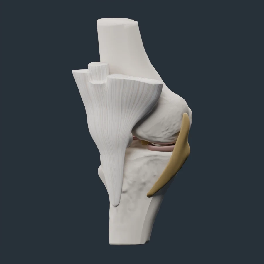
Anatomy at a Glance
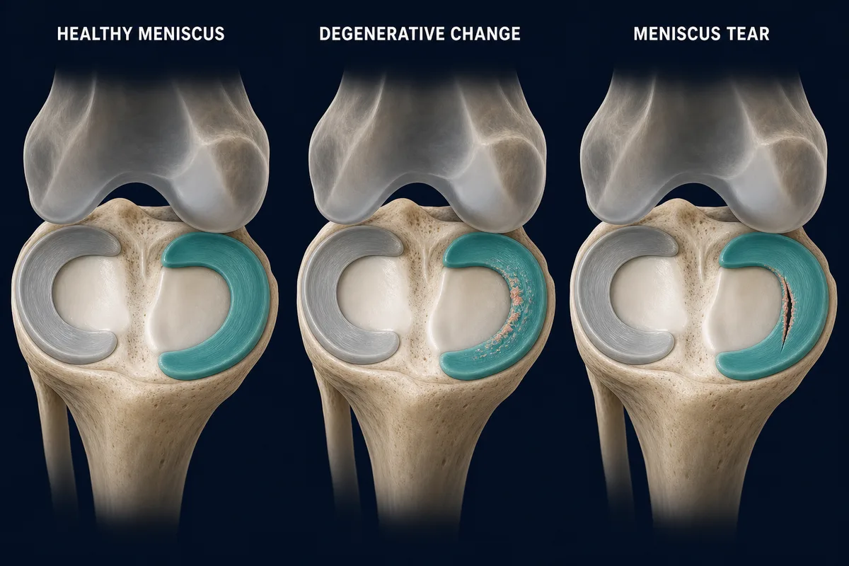
Meniscal changes range from gradual fraying to a defined tear. The appearance of a tear does not by itself determine whether it is painful or needs surgery.
Overview
A meniscus tear affects one of the two fibrocartilage pads between the thighbone and shinbone. Tears may happen suddenly during a loaded twist or develop gradually as the tissue changes over time.
An MRI tear is not automatically the cause of knee pain. Meniscal changes are common, particularly with age, and must be interpreted alongside the injury history, symptoms and examination.
Common Clues
- Pain focused at the inner or outer joint line
- Swelling that may build over several hours
- Pain with loaded twisting or deep bending
- Catching or a sense that movement is not smooth
- Difficulty fully straightening the knee
Clicking alone does not prove a tear. True locking means the knee becomes mechanically stuck and cannot fully straighten, rather than merely feeling painful or stiff.
Tear Type Matters
Treatment is influenced by whether the tear is traumatic or degenerative, its shape and location, whether it is displaced, tissue quality, blood supply and associated ACL or cartilage injury. Root tears and displaced bucket-handle tears deserve particular attention because they can substantially change meniscus function or block motion.
Treatment
Many non-displaced tears and degenerative meniscus symptoms begin with non-surgical care: activity modification, restoring motion and progressive lower-limb strengthening. Surgery is not automatically better simply because MRI shows a tear.
Repair may be considered when the tear pattern and tissue are suitable, especially when preserving meniscus function is possible. Partial removal is generally reserved for selected symptomatic tissue that cannot be repaired. The decision is individualized.
When to Get Assessed
Arrange assessment after a traumatic twist with swelling, persistent joint-line pain, instability or catching. A knee that is truly locked, a major injury with inability to bear weight, deformity or marked swelling needs prompt care.
Related Reading
References
- Pujol N, et al. “The Formal EU-US Meniscus Rehabilitation 2024 Consensus: Part II—Prevention, Nonoperative Treatment and Return to Sport.” https://pubmed.ncbi.nlm.nih.gov/40519541/
- AAOS. “Acute Isolated Meniscal Pathology Clinical Practice Guideline.” https://www.aaos.org/quality/quality-programs/lower-extremity-programs/acute-isolated-meniscal-pathology/
This information is for education only and is not a medical diagnosis. InjuryAtlas does not replace an assessment by a qualified health professional. If you have severe pain, visible deformity, numbness, loss of function, or symptoms that are getting worse, seek medical care promptly.