Overview
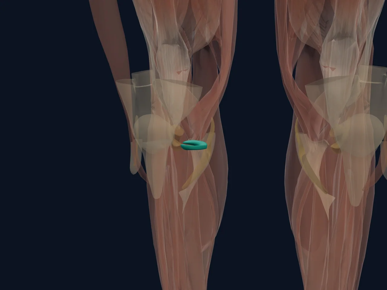
The medial meniscus is the crescent-shaped pad of fibrocartilage between the femur and tibia on the inside of the knee. It spreads load, improves joint fit, absorbs shock and contributes to stability and joint awareness.
Location and Attachments
- Sits on the medial tibial plateau
- Has anterior and posterior horns anchored to the tibia
- Attaches to the capsule and deep fibres of the MCL
- Is less mobile than the lateral meniscus
What It Does
The meniscus turns compressive load into circumferential “hoop” stress and distributes force over a broader joint surface. It also contributes to lubrication, proprioception and stability. Preserving functioning meniscal tissue matters because removing it increases contact stress on articular cartilage.
Blood Supply and Healing
The outer edge has the best blood supply. The inner portion relies largely on diffusion from joint fluid. Tear location is therefore one factor in healing potential, but pattern, stability, age, tissue quality and associated injuries also matter.
Common Problems
- Acute tear during loaded rotation
- Degenerative tearing that develops over time
- Root tear near a horn attachment
- Injury occurring with an ACL tear
Clicking or an MRI finding does not automatically mean the meniscus is the pain source. Some tears cause few or no symptoms.
Clinical Relevance
Persistent locking, inability to fully straighten the knee, major swelling or symptoms after a traumatic twist should be assessed. Diagnosis combines history, examination and imaging when it will change management.
Related Anatomy
- Meniscus rehabilitation guide
- Lateral meniscus
- Medial collateral ligament
- Anterior cruciate ligament
References
- StatPearls. "Anatomy, Bony Pelvis and Lower Limb: Medial Meniscus." https://www.ncbi.nlm.nih.gov/books/NBK537276/
- StatPearls. "Knee Meniscal Tears." https://www.ncbi.nlm.nih.gov/books/NBK431067/