Overview
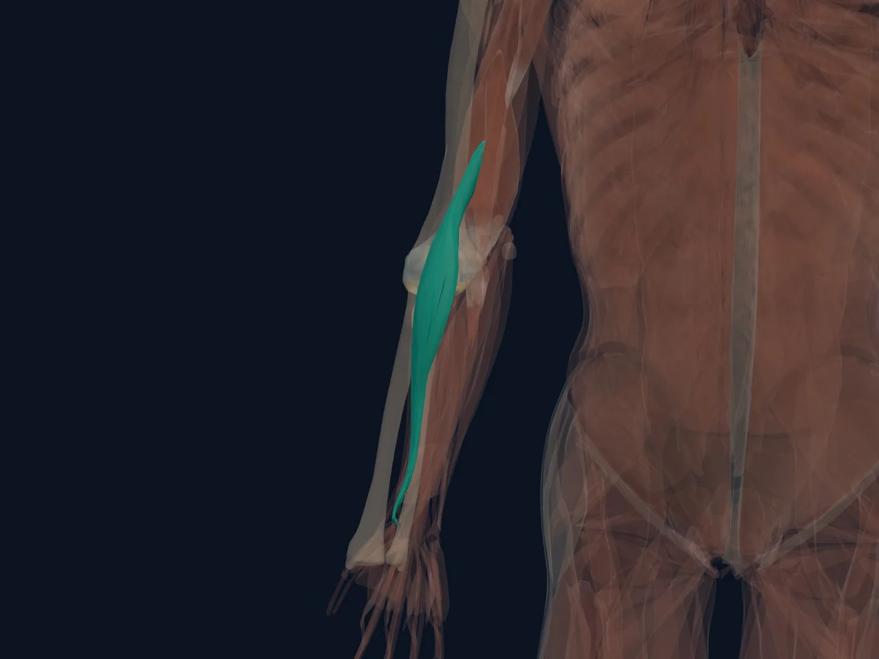
The brachioradialis is the prominent muscle along the thumb side of the forearm. Despite living mostly in the forearm, its main job is to bend the elbow.
It works especially well when the forearm is in a neutral, thumb-up position—the grip used for a hammer curl, carrying a suitcase or lifting a mug. It can also help bring the forearm back toward neutral after the palm has turned fully up or down.
Location
- Begins on the outer lower humerus above the elbow
- Crosses the elbow without crossing the wrist
- Runs down the thumb side of the forearm to the radius
Origin, Insertion, Nerve Supply, and Action
- Origin: Proximal two-thirds of the lateral supracondylar ridge of the humerus
- Insertion: Lateral distal radius near the styloid process
- Nerve supply: Radial nerve, mainly C5–C7
- Action: Flexes the elbow most effectively with the forearm in neutral
Function
- Bends the elbow during neutral-grip lifting
- Helps return the forearm toward neutral from full pronation or supination
- Assists the biceps and brachialis during carrying and pulling
Common Problems
The brachioradialis can become sore after a sharp increase in hammer curls, climbing, gripping or repetitive tool use. Pain is often felt near the outer elbow or along the upper thumb-side forearm.
That location overlaps with tennis elbow, radial-nerve irritation and other forearm muscles. A tender spot or painful curl cannot diagnose the source on its own.
When to Get It Assessed
Arrange an assessment if pain follows a sudden injury, grip or wrist strength is clearly reduced, numbness or tingling develops, or symptoms continue despite reducing the aggravating load.
Related Anatomy
References
- OpenStax. “Muscles of the Pectoral Girdle and Upper Limbs.” https://openstax.org/books/anatomy-and-physiology-2e/pages/11-5-muscles-of-the-pectoral-girdle-and-upper-limbs
- Moore KL et al. Clinically Oriented Anatomy. Wolters Kluwer.
- Standring S, ed. Gray’s Anatomy: The Anatomical Basis of Clinical Practice. Elsevier.