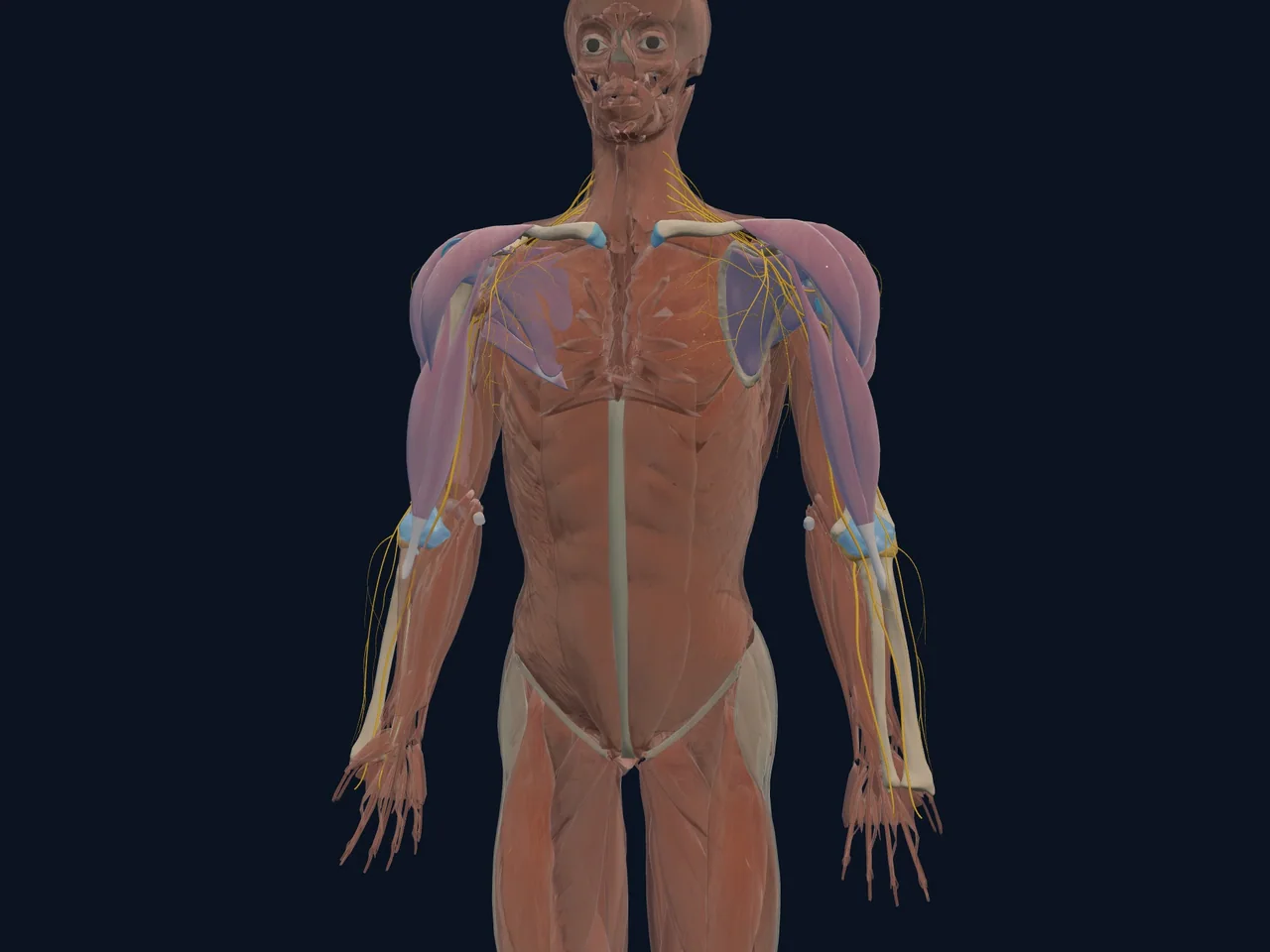
Overview
Your shoulder relies on a network of tough, rope-like ligaments — bands of tissue that connect bone to bone — to add stability that the shallow shoulder socket can't provide on its own. Unlike muscles, ligaments don't actively contract; they act more like static ropes or straps that stop joints from moving too far in a given direction. Because the shoulder has such a huge range of motion, it needs several distinct ligaments working in different areas to prevent it from moving too far and slipping out of place.
Location
- Glenohumeral ligaments (superior, middle, and inferior bands) — thicken parts of the shoulder joint capsule itself, running between the humerus and the rim of the glenoid socket
- Coracoacromial ligament — spans between the coracoid process and the acromion, forming a protective arch over the top of the shoulder joint
- Coracoclavicular ligaments (conoid and trapezoid) — run from the coracoid process of the shoulder blade up to the underside of the collarbone
- Acromioclavicular ligaments — directly reinforce the small AC joint at the top of the shoulder
Function
- Glenohumeral ligaments resist excessive rotation and translation (sliding) of the humeral head within the socket, particularly during extreme positions like the arm raised and rotated outward
- The coracoacromial ligament, along with the acromion and coracoid process, forms a protective bony-ligamentous arch over the top of the rotator cuff tendons
- Coracoclavicular ligaments provide the primary vertical stability for the collarbone, preventing it from shifting upward relative to the shoulder blade
- Acromioclavicular ligaments provide direct stability to the small joint at the top of the shoulder
Structures It Connects To
- Humerus (upper arm bone) and scapula (shoulder blade) — connected by the glenohumeral ligaments
- Coracoid process and acromion (both parts of the scapula) — connected by the coracoacromial ligament
- Coracoid process and clavicle (collarbone) — connected by the coracoclavicular ligaments
- Clavicle and acromion — connected directly by the acromioclavicular ligaments
- Rotator cuff tendons and subacromial bursa — sit just beneath the coracoacromial ligament's protective arch
Movements It Assists
Ligaments don't actively move joints the way muscles do — instead, they define the outer limits of safe movement, allowing full range of motion while preventing the joint from going too far:
- Glenohumeral ligaments limit excessive external rotation and forward/backward sliding of the humeral head
- Coracoclavicular ligaments limit excessive upward movement of the collarbone relative to the shoulder blade
- Acromioclavicular ligaments limit excessive movement at the AC joint in multiple directions
- The coracoacromial ligament, together with nearby bone, defines the available space for the rotator cuff tendons to glide through
Common Injuries
- Shoulder dislocation — often involves stretching or tearing of the glenohumeral ligaments, especially the inferior band, which is the most commonly damaged in forward (anterior) dislocations
- Acromioclavicular joint injury (AC sprain/separation) — involves the acromioclavicular and, in more severe cases, the coracoclavicular ligaments
- Shoulder impingement syndrome — can be influenced by a thickened or irritated coracoacromial ligament reducing the space for the rotator cuff
- Chronic shoulder instability — repeated stretching of the glenohumeral ligaments over time can make the joint more prone to repeat dislocation
Pain Referral Patterns
- Glenohumeral ligament injuries typically cause deep, aching pain felt throughout the shoulder, often worse with the arm in the position that caused the injury (commonly overhead and rotated outward)
- Coracoclavicular and acromioclavicular ligament injuries cause sharp, localized pain right at the top of the shoulder
- Ligament-related instability often comes with a sensation of the shoulder feeling like it might "give way" or slip, rather than a constant ache
Clinical Relevance
Because ligaments have a limited blood supply compared to muscle, they generally heal more slowly, and severe ligament injuries (like a complete AC joint separation with torn coracoclavicular ligaments) sometimes need surgical repair rather than relying on natural healing alone (StatPearls: Acromioclavicular Joint Injury). For shoulder dislocations, clinicians assess how much ligament laxity or damage is present, since repeated stretching of the glenohumeral ligaments after a first dislocation is a major reason some people go on to experience recurring instability.
Interesting Facts
- The coracoacromial ligament, together with the bones it connects, is sometimes called the shoulder's "roof" — and a thickened or overly tight version of this ligament can narrow the space available for the rotator cuff tendons underneath it.
- The inferior glenohumeral ligament is considered the single most important stabilizing structure against the shoulder dislocating forward, especially when the arm is raised and rotated outward — a position often described as the "throwing position."
- Unlike the knee's well-known ACL and MCL, the shoulder's ligaments work more as a group effort, since no single shoulder ligament provides as much stability on its own as some of the major knee ligaments do.
Related Anatomy
- Glenohumeral joint — stabilized primarily by the glenohumeral ligaments
- Acromioclavicular joint — stabilized by the acromioclavicular and coracoclavicular ligaments
- Scapula — the coracoid process and acromion, both part of the scapula, anchor several of these ligaments
- Clavicle — anchored from below by the coracoclavicular ligaments
- Subacromial bursa — sits just beneath the coracoacromial ligament arch
Related Injuries
References
- StatPearls (NCBI Bookshelf). "Acromioclavicular Joint Injury." https://www.ncbi.nlm.nih.gov/books/NBK493188/
- Physiopedia. "Glenohumeral Joint." https://www.physio-pedia.com/Glenohumeral_Joint
- Kenhub. "Ligaments of the shoulder joint." https://www.kenhub.com/en/library/anatomy/ligaments-of-the-shoulder-joint
- PMC. "Anatomy and Biomechanics of the Shoulder." https://pmc.ncbi.nlm.nih.gov/articles/PMC5039561/