Overview
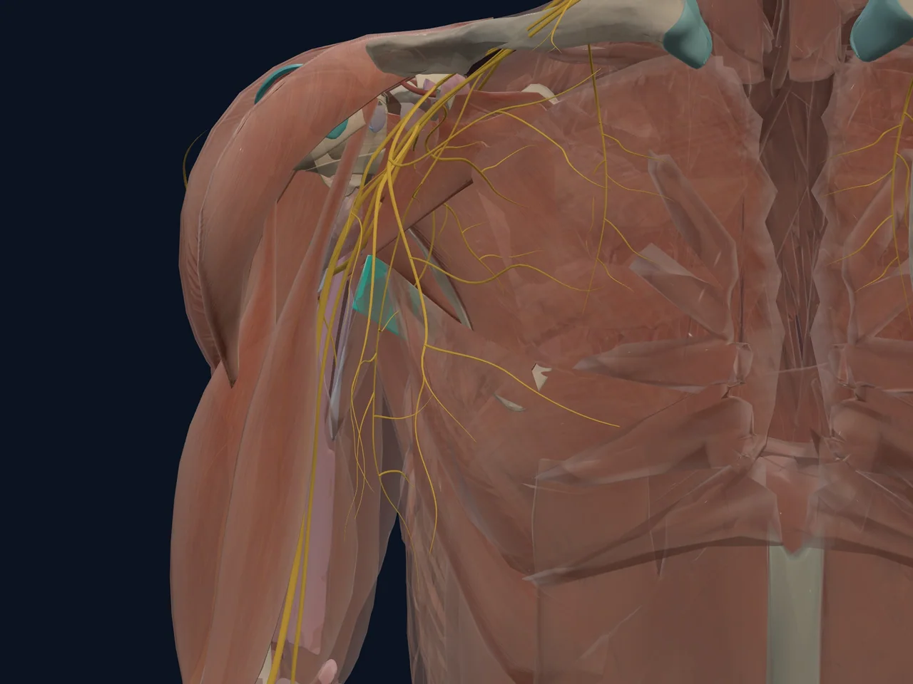
The teres major is a thick, strap-like muscle running from the bottom corner of your shoulder blade to the front of your upper arm bone. Despite sitting right next to the rotator cuff on the back of the shoulder, it is not a rotator cuff muscle — it does not blend into the joint capsule, and its role is to move the arm rather than fine-tune the ball in the socket. It works so closely with the much larger latissimus dorsi that the two are often described as a functional pair.
Location
- Arises from the lower, outer corner of the scapula (shoulder blade)
- Runs upward and outward across the back of the shoulder to the front of the humerus (upper arm bone)
- Forms the lower border of the posterior axillary fold you can feel at the back of your armpit
Origin, Insertion, Nerve Supply, and Action
- Origin: Dorsal surface of the inferior angle and lower lateral border of the scapula
- Insertion: Medial lip of the intertubercular (bicipital) groove of the humerus
- Nerve supply: Lower subscapular nerve (spinal nerve roots C5–C6, sometimes C7)
- Action: Adduction, internal (medial) rotation, and extension of the arm at the shoulder
In plain English: the teres major helps pull your arm down, back and inward — the motion you use when rowing, swimming or pushing yourself up out of a chair.
Function
- Adducts the arm, bringing it back toward the body
- Internally rotates the shoulder
- Extends the arm at the shoulder from a flexed position
- Works alongside the latissimus dorsi during pulling and pushing-off movements
Structures It Connects To
- Scapula (shoulder blade) — its origin at the inferior angle
- Humerus (upper arm bone) — its insertion on the medial lip of the bicipital groove
- Teres minor — a separate, smaller muscle just above it with the opposite action (external rotation)
- Subscapularis — shares the internal-rotation role from the front of the shoulder blade
Movements It Assists
- Adduction of the shoulder
- Internal rotation of the shoulder
- Extension of the shoulder during pulling movements
Common Injuries
- Teres major strain — usually an overuse or sudden-overload injury in throwing, swimming and climbing athletes
- Teres major tendinopathy — irritation at the humeral attachment with repetitive loading
- Rotator cuff tear — the teres major is sometimes evaluated alongside the cuff, though it is a distinct, non-cuff muscle
Pain Referral Patterns
- Aching or tightness at the back of the shoulder near the lower edge of the shoulder blade
- Pain when reaching up and forward, or when strongly pulling the arm down and in
- Soreness felt during the follow-through phase in throwing and racquet athletes
Clinical Relevance
Because the teres major is a mover rather than a stabiliser, it tends to be a source of strain and overuse soreness rather than the tendon tears seen in the rotator cuff. In throwing athletes, teres major and latissimus dorsi injuries are increasingly recognised as a cause of posterior arm and axillary pain that can sideline a season if not managed with graded loading.
Interesting Facts
- Its name comes from Latin: "teres" means rounded or cylindrical, and "major" distinguishes it from the smaller teres minor nearby — the two share a name but have opposite actions.
- Together with latissimus dorsi, it forms part of the muscular border of the posterior wall of the armpit.
Related Anatomy
- Teres minor — the smaller neighbour with the opposite (external-rotation) action
- Subscapularis — another internal rotator of the shoulder
- Scapula — the bone the teres major originates from
- Humerus (proximal) — the bone it inserts on
Related Injuries
References
- Physiopedia. "Teres Major." https://www.physio-pedia.com/Teres_Major
- Kenhub. "Teres major muscle: Anatomy, course and function." https://www.kenhub.com/en/library/anatomy/teres-major-muscle
- Terminologia Anatomica (FIPAT). https://fipat.library.dal.ca/