Overview
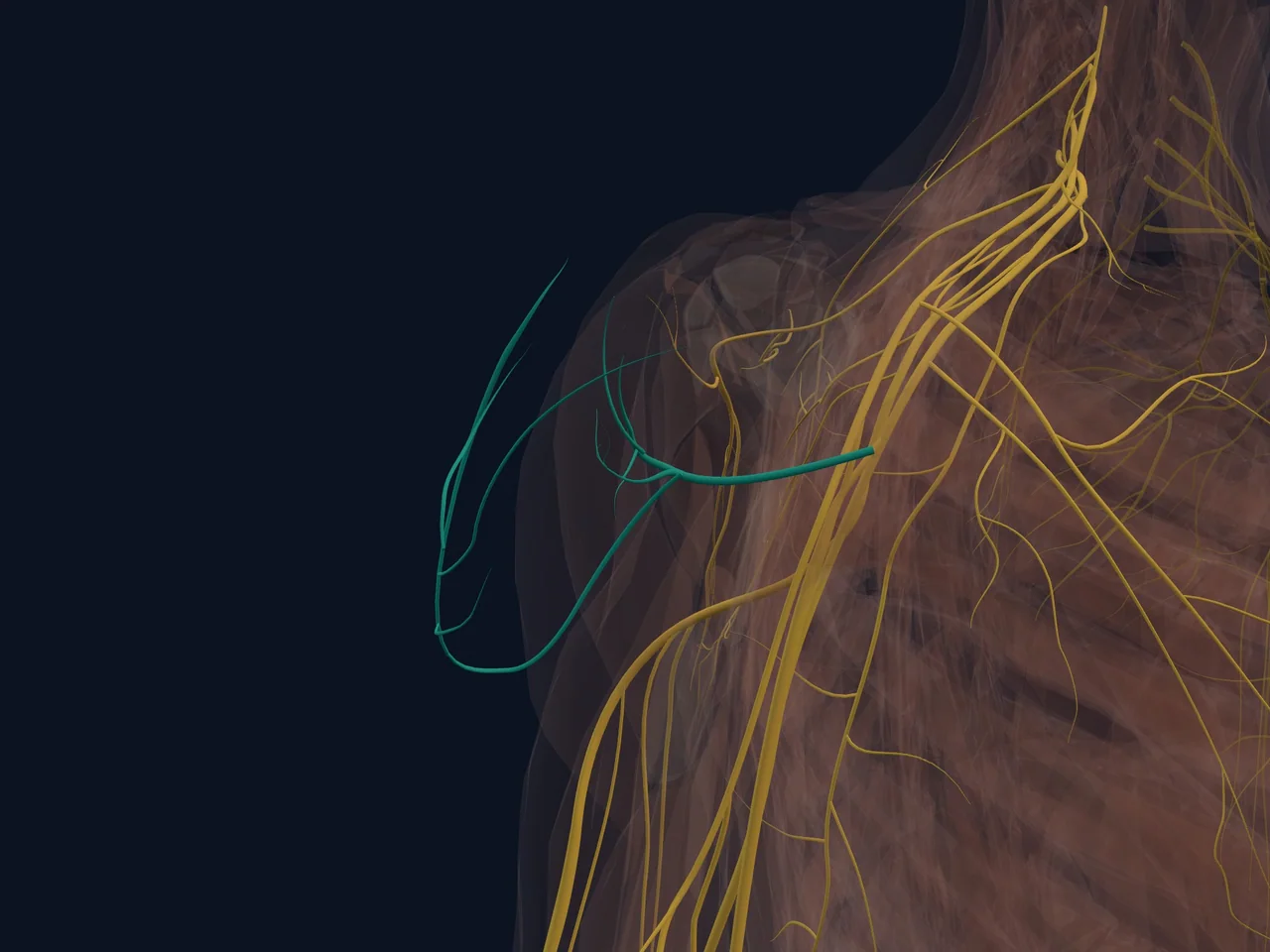
The axillary nerve (sometimes called the circumflex nerve) is the nerve that powers your deltoid, the big muscle that caps your shoulder and lets you lift your arm out to the side. It travels close to the shoulder joint itself, curving around the back of your upper arm bone, which puts it at risk any time the shoulder joint is forcefully injured — most notably during a shoulder dislocation. When this nerve is stretched or damaged, the result is often a weak, flattened-looking shoulder because the deltoid can't do its job properly.
Location
- Branches off the posterior cord of the brachial plexus, the network of nerves that supplies the arm, carrying fibers mainly from the C5 and C6 spinal nerve roots
- Travels behind the axillary artery and in front of the subscapularis muscle, deep in the armpit (axilla) region
- Passes through the quadrangular space, a small gap bordered by the teres minor, teres major, the long head of triceps, and the upper arm bone
- Curves around the back of the surgical neck of the humerus (upper arm bone), sitting directly against the bone at this point
Function
- Provides motor (movement) signals to the deltoid muscle, allowing it to lift the arm out to the side and overhead
- Provides motor signals to the teres minor muscle, assisting external rotation of the arm
- Supplies sensation to a small patch of skin over the lower, outer part of the deltoid — sometimes called the "regimental badge area," because it corresponds roughly to where a military rank badge sits on a uniform sleeve
- Contributes sensory branches to the glenohumeral (shoulder socket) joint capsule
In plain English: this nerve is the "wiring" for your shoulder-cap muscle. If it's damaged, your deltoid can't contract properly, which makes lifting your arm out to the side difficult even though the muscle itself is healthy (Cleveland Clinic: Axillary Nerve).
Structures It Connects To
- Brachial plexus — its origin, specifically the posterior cord
- Quadrangular space — the anatomical passage this nerve travels through on its way to the back of the shoulder
- Deltoid muscle — its main motor target
- Teres minor muscle — also receives motor branches from this nerve
- Surgical neck of the humerus — the nerve wraps directly around this part of the bone, making it vulnerable during fractures here
- Glenohumeral joint capsule — receives sensory branches from this nerve
Movements It Assists
- Shoulder abduction (raising the arm out to the side) — via its supply to the deltoid
- Shoulder flexion and extension, partially — also via the deltoid's front and back fibers
- External rotation of the arm — via its supply to the teres minor
Common Injuries
- Axillary nerve injury/palsy — most commonly caused by shoulder dislocation, since the nerve sits so close to the joint capsule and can be stretched when the humeral head moves out of place
- Proximal humerus fracture — because the nerve wraps directly around the surgical neck of the humerus, fractures in this area can stretch or injure the nerve
- Quadrangular space syndrome — a rare condition where the nerve is compressed within the quadrangular space, sometimes seen in overhead throwing athletes
Pain Referral Patterns
- Axillary nerve injury more often causes numbness and weakness than pain on its own, though it frequently occurs alongside a painful injury like a dislocation
- Numbness is typically felt over the lower, outer part of the shoulder and upper arm (the "regimental badge area")
- Weakness specifically with lifting the arm out to the side, despite the deltoid muscle itself being structurally intact, is the hallmark clinical sign
Clinical Relevance
Checking axillary nerve function is a standard part of assessing any shoulder dislocation, both before and after the joint is put back into place — clinicians test deltoid strength and sensation over the "regimental badge area" specifically because this nerve is the most commonly injured nerve in shoulder dislocations (Cleveland Clinic: Axillary Nerve). Most axillary nerve stretch injuries improve gradually over weeks to months as the nerve heals, though recovery time varies and more severe injuries sometimes need further specialist evaluation.
Interesting Facts
- The patch of skin this nerve supplies sensation to is nicknamed the "regimental badge area" because it roughly matches where a rank badge would sit on a military uniform's short sleeve.
- Because the axillary nerve hugs the bone so closely around the surgical neck of the humerus, this specific fracture location is a classic teaching example of a nerve injury linked directly to a bone injury.
- Despite supplying a large, powerful muscle (the deltoid), the axillary nerve itself is a relatively small nerve — a good example of how nerve size doesn't necessarily reflect the size of the muscle it controls.
Related Anatomy
- Deltoid — the main muscle supplied by this nerve
- Teres minor — also supplied by this nerve
- Glenohumeral joint — this nerve runs close to the joint capsule and is vulnerable during dislocation
- Proximal humerus — the nerve wraps around the surgical neck of this bone
- Brachial plexus (shoulder overview) — the nerve network this nerve branches from
Related Injuries
- Shoulder dislocation
- Proximal humerus fracture — this nerve wraps closely around the fracture-prone surgical neck of the humerus
References
- StatPearls (NCBI Bookshelf). "Anatomy, Shoulder and Upper Limb, Axillary Nerve." https://www.ncbi.nlm.nih.gov/books/NBK493212/
- StatPearls (NCBI Bookshelf). "Anatomy, Shoulder and Upper Limb, Axilla." https://www.ncbi.nlm.nih.gov/books/NBK547723/
- Cleveland Clinic. "Axillary Nerve: Overview, Function & Anatomy." https://my.clevelandclinic.org/health/body/22264-axillary-nerve
- Kenhub. "Axillary nerve: Anatomy, course, function." https://www.kenhub.com/en/library/anatomy/axillary-nerve
- Physiopedia. "Axillary Nerve." https://www.physio-pedia.com/Axillary_Nerve